
Title: “Urology Uncovered: Your Complete Guide to a Healthy Urinary System & What Every Patient Should Know”

—

Introduction – Why the “Little‑Talk” About Urology Matters

When you hear the word urology, most people picture a sterile exam room, a white coat, and a brief, uncomfortable conversation about “that thing down there.” Yet the urinary system is the body’s hidden powerhouse – it filters waste, balances fluids, regulates blood pressure, and even influences sexual health. Ignoring it can lead to painful kidney stones, embarrassing incontinence, or life‑threatening cancers that are often preventable with early detection.

In this 2,000‑word deep dive, we’ll demystify urology, break down the most common conditions, and arm you with actionable steps to keep your kidneys, bladder, prostate, and urethra in top shape. Whether you’re a patient, a caregiver, or simply health‑curious, this guide will give you the confidence to talk openly with your urologist and take charge of your urinary health.

—

1. The Foundations of Urology: Anatomy, Function, and Why It All Matters

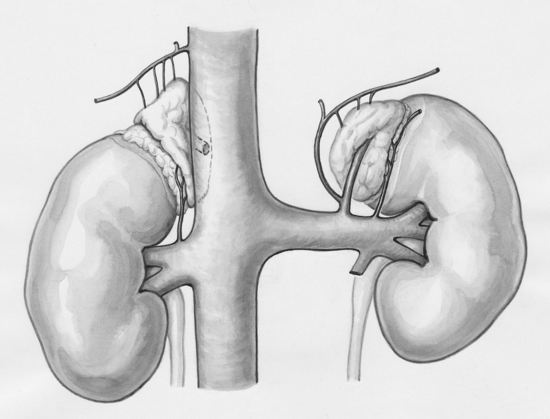
1.1 The Urinary System – A Quick Anatomy Crash Course

| Organ | Primary Role | Key Health Indicators |
|——-|————–|———————–|
| Kidneys | Filter blood, remove waste, regulate electrolytes & blood pressure | Creatinine, eGFR, urine output |
| Ureters | Transport urine from kidneys to bladder | Obstruction signs (pain, hydronephrosis) |
| Bladder | Stores urine until convenient elimination | Capacity, residual volume |
| Urethra | Conduit for urine (and semen in males) | Flow rate, continence |
| Prostate (men) | Produces seminal fluid, surrounds urethra | PSA level, size, urinary flow |

Understanding where each part sits helps you recognize symptoms early. For example, flank pain often signals kidney involvement, while a weak stream points to bladder or prostate issues.

1.2 How the System Works Together – The “Filtration‑Storage‑Elimination” Cycle

1. Filtration: Blood enters the kidneys where nephrons extract waste and excess fluid.
2. Reabsorption: Essential nutrients, water, and electrolytes are returned to the bloodstream.
3. Excretion: The filtered urine travels down the ureters into the bladder, where it’s stored.
4. Elimination: When the bladder reaches ~300–500 ml, nerves signal the brain, prompting a coordinated muscle contraction that pushes urine through the urethra.

Disruption at any stage can cause symptoms that range from subtle (frequent nighttime urination) to severe (acute renal failure).

1.3 Why Urology Is a Lifelong Specialty

Urology isn’t just about “men’s health” or “kidney stones.” It covers:

  • Pediatric urology: Congenital anomalies like vesicoureteral reflux.
  • Female urology: Stress incontinence, pelvic organ prolapse, and recurrent urinary tract infections (UTIs).
  • Oncologic urology: Bladder, kidney, and prostate cancers.
  • Reconstructive urology: Post‑surgical repairs, urinary diversions, and gender‑affirming procedures.
  • Because the urinary system interacts with the cardiovascular, endocrine, and reproductive systems, a urologist often collaborates with other specialists to deliver holistic care.

    —

    2. Common Urologic Conditions & How to Spot Them Early

    2.1 Kidney Stones – The Painful Pebbles

    Key facts:

  • Prevalence: ~1 in 10 people will develop a stone in their lifetime.
  • Types: Calcium oxalate (most common), uric acid, struvite, cystine.
  • Red‑flag symptoms:

  • Sudden, severe flank pain radiating to the groin (often described as “one of the worst pains”).
  • Hematuria (blood in urine).
  • Nausea, vomiting, or fever (suggests infection).
  • Actionable steps:

    | Step | What to Do | Why It Helps |
    |——|————|————–|
    | Hydration | Aim for 2–3 L of water daily (clear urine). | Dilutes urine, reduces crystal formation. |
    | Diet tweak | Limit high‑oxalate foods (spinach, nuts), moderate animal protein, reduce sodium. | Lowers calcium oxalate supersaturation. |
    | Medical evaluation | Get a non‑contrast CT scan if pain is acute; a stone analysis if you pass one. | Guides targeted prevention (e.g., potassium citrate for uric acid stones). |
    | Medication | Thiazide diuretics for calcium stones; allopurinol for uric acid stones. | Alters urinary chemistry to prevent recurrence. |

    When to call a urologist: Persistent pain > 24 hrs, fever > 38°C, or inability to pass urine. Early intervention can prevent renal damage and the need for invasive surgery.

    2.2 Urinary Tract Infections (UTIs) – The “Burning” Problem

    Who gets them? Women (especially sexually active), the elderly, and individuals with catheters.

    Typical signs:

  • Burning sensation during urination.
  • Cloudy, foul‑smelling urine.
  • Pelvic pressure or lower back pain.
  • Quick self‑care checklist:

    1. Increase fluid intake – 1.5–2 L of water daily to flush bacteria.
    2. Cranberry products – May reduce bacterial adhesion (evidence modest).
    3. Proper hygiene – Front‑to‑back wiping, urinating after intercourse.

    Medical management:

  • First‑line antibiotics (e.g., nitrofurantoin, trimethoprim‑sulfamethoxazole) for 3–5 days.
  • Culture‑guided therapy for recurrent cases or complicated UTIs.
  • Red flag: Fever, flank pain, or confusion (especially in seniors) may indicate a kidney infection (pyelonephritis) requiring urgent care.

    2.3 Benign Prostatic Hyperplasia (BPH) – “Aging” Prostate

    Prevalence: Affects > 50 % of men over 60.

    Symptoms (the “LUTS” – lower urinary tract symptoms):

  • Storage issues: Frequent urination, nocturia, urgency.
  • Voiding issues: Weak stream, hesitancy, incomplete emptying.
  • Lifestyle hacks:

  • Timed voiding: Schedule bathroom trips every 2–3 hrs to train bladder capacity.
  • Limit bladder irritants: Caffeine, alcohol, carbonated drinks.
  • Pelvic floor exercises: Strengthen muscles that aid bladder emptying.
  • Medical options:

    | Option | Mechanism | Typical Candidates |
    |——–|———–|——————–|
    | Alpha‑blockers (tamsulosin) | Relax prostate smooth muscle → improved flow. | Moderate symptoms, no large prostate. |
    | 5‑alpha‑reductase inhibitors (finasteride) | Shrink prostate over months. | Larger prostates (>30 g) or combination therapy. |
    | Minimally invasive procedures (UroLift, Rezūm) | Physical remodeling of tissue. | Men seeking surgery‑free relief. |

    When to see a urologist: Persistent nocturia (>2‑3 times/night), urinary retention, or recurrent UTIs. Early evaluation can prevent bladder damage and kidney back‑pressure.

    2.4 Urinary Incontinence – Regaining Control

    Types:

  • Stress incontinence: Leakage with coughing, sneezing, or lifting.
  • Urgency incontinence: Sudden urge, often due to overactive bladder.
  • Mixed incontinence: Combination of both.
  • First‑line strategies:

    1. Kegel exercises – Contract pelvic floor for 5 seconds, relax 5 seconds; 10 reps × 3 sets daily.
    2. Bladder training – Delay voiding by 5‑10 minutes, gradually extending intervals.
    3. Weight management – Reducing BMI lessens pressure on pelvic floor.

    Medical & device options:

  • Anticholinergics (oxybutynin) for urgency.
  • Beta‑3 agonists (mirabegron) – fewer dry‑mouth side effects.
  • Urethral slings or bulking agents for stress incontinence (surgical).
  • Red flag: Sudden onset of incontinence with fever, flank pain, or neurological changes may signal spinal cord issues or infection—seek immediate care.

    2.5 Urologic Cancers – Early Detection Saves Lives

    | Cancer | Screening Tool | Typical Age | Key Symptom |
    |——–|—————-|————|————-|
    | Prostate | PSA blood test + DRE | 50 + (earlier if high risk) | Difficulty urinating, blood in urine |
    | Bladder | Urine cytology, cystoscopy (high‑risk) | 55 + | Hematuria (painless) |
    | Kidney | Imaging (ultrasound/CT) for incidental findings | 45 + | Flank mass, hematuria |
    | Testicular | Self‑exam | 15‑35 | Lump or heaviness in scrotum |

    Actionable prevention checklist:

  • Quit smoking – Reduces bladder and kidney cancer risk by up to 50 %.
  • Stay hydrated – Dilutes carcinogens in urine.
  • Balanced diet – High fruit/vegetable intake, limited processed meats.
  • Regular check‑ups – Discuss PSA testing with your doctor if you have a family history.
  • —

    3. Lifestyle & Home‑Based Strategies for Optimal Urinary Health

    3.1 Hydration – The Gold Standard

  • Goal: 2–2.5 L of fluid daily for most adults; adjust for climate, activity level, and kidney disease.
  • Best choices: Water, herbal teas, low‑sugar fruit infusions.
  • Limit: Sugary sodas, excessive caffeine, and high‑salt sports drinks (they can increase stone risk and irritate the bladder).
  • Tip: Carry a reusable bottle with volume markers; sip consistently rather than gulping large amounts at once.

    3.2 Nutrition Tailored to the Urinary Tract

    | Nutrient | Sources | Urology Benefit |
    |———-|———|—————–|
    | Calcium (moderate) | Low‑fat dairy, fortified plant milks | Binds oxalate in gut, reduces stone formation. |
    | Potassium citrate | Citrus fruits, berries | Raises urine pH, preventing uric acid stones. |
    | Omega‑3 fatty acids | Salmon, flaxseed | Anti‑inflammatory; may lower prostate cancer progression. |
    | Fiber | Whole grains, legumes | Improves bowel health, reduces pressure on bladder. |

    Avoid: Excessive animal protein (increases calcium and uric acid excretion) and high‑salt diets (raises calcium in urine).

    3.3 Exercise & Pelvic Floor Strengthening

  • Aerobic activity (30 min brisk walk, 5 days/week) improves circulation, lowers blood pressure, and reduces stone risk.
  • Pelvic floor training – Use biofeedback apps or a pelvic floor therapist for correct technique.
  • Weight‑bearing resistance – Helps maintain muscle tone that supports bladder and prostate health.
  • 3.4 Smart Bathroom Habits

    1. Don’t “hold it” – Chronic retention can stretch bladder walls and cause overflow incontinence.
    2. Double void – After finishing, wait a minute and try again to empty residual urine.
    3. Proper positioning – For men, standing can aid complete emptying; for women, a relaxed seated posture reduces pelvic floor strain.

    3.5 When to Use Over‑the‑Counter (OTC) Aids

  • Cranberry capsules – May prevent recurrent UTIs in women with a history of ≥ 3 infections per year.
  • Phenazopyridine – Short‑term (≤ 2 days) pain relief for urinary discomfort; not a cure.
  • Lactobacillus probiotic – Supports healthy vaginal flora, indirectly reducing UTI risk.
  • Caution: Always discuss OTC use with your urologist, especially if you have kidney disease or are on prescription diuretics.

    —

    4. Navigating the Urology Appointment – What to Expect and How to Prepare

    4.1 Choosing the Right Urologist

  • Board certification – Verify through the American Board of Urology (or your country’s equivalent).
  • Specialty focus – Pediatric, female, oncologic, or reconstructive urology.
  • Patient reviews & communication style – A doctor who explains procedures in plain language improves adherence.

4.2 Pre‑Visit Checklist

| Item | Why It Matters |
|——|—————-|
| Medication list | Some drugs (e.g., diuretics, anticoagulants) affect urine tests. |
| Symptom diary (frequency, pain,

Title: “Beyond the Bladder: A Complete Guide to Benign Prostatic Hyperplasia (BPH) – Causes, Symptoms, Treatments & Everyday Tips for Prostate Health”

—

Introduction – Why the “Silent” Enlargement Deserves Your Attention

Imagine waking up at 2 a.m. to rush to the bathroom, only to discover you’ve been up for an hour with no relief. Or picture yourself at a social gathering, constantly scanning the room for the nearest restroom. For millions of men over 40, this isn’t a one‑off nightmare—it’s a daily reality caused by benign prostatic hyperplasia (BPH), more commonly known as an enlarged prostate.

Despite the name, BPH isn’t cancerous. Yet its impact on quality of life can be profound, affecting sleep, work productivity, and even emotional wellbeing. The good news? With the right knowledge, you can spot the early warning signs, get an accurate diagnosis, and choose a treatment plan that fits your lifestyle.

In this 2,000‑word deep dive we’ll explore:

  • What benign prostatic hyperplasia really is and why it happens.
  • The most common BPH symptoms and how to differentiate them from other prostate issues.
  • Proven diagnostic tools and when to seek professional help.
  • A spectrum of treatment options—from medication and minimally invasive procedures to lifestyle tweaks that can ease urinary problems.
  • Practical, actionable tips for everyday prostate health.
  • Let’s demystify BPH together and empower you to take control of your urinary health—starting right now.

    —

    1. Understanding Benign Prostatic Hyperplasia – The Basics

    1.1 What Is BPH?

    The prostate is a walnut‑sized gland located just below the bladder, surrounding the urethra (the tube that carries urine out of the body). Benign prostatic hyperplasia refers to the non‑cancerous enlargement of this gland. As the prostate grows, it can compress the urethra, leading to the classic urinary complaints many men experience.

    Key facts at a glance

    | Fact | Detail |
    |——|——–|
    | Prevalence | Affects ~50 % of men by age 60 and up to 90 % by age 85 |
    | Onset | Typically begins in the early 40s, symptoms often appear later |
    | Cause | Multifactorial: hormonal changes (especially dihydrotestosterone), aging, inflammation, and genetics |
    | Not Cancer | “Benign” means it does not turn into prostate cancer, though both can coexist |

    1.2 Why Does the Prostate Grow?

    While the exact trigger isn’t fully understood, researchers agree on a few major contributors:

    1. Hormonal Shifts – As men age, testosterone gradually declines while estrogen levels stay relatively stable. The balance tips toward dihydrotestosterone (DHT), a potent androgen that stimulates prostate cell proliferation.
    2. Growth Factors – Proteins like insulin‑like growth factor‑1 (IGF‑1) and fibroblast growth factor (FGF) can promote tissue expansion.
    3. Inflammation – Chronic prostatitis or subclinical inflammation may act as a “fuel” for cellular growth.
    4. Genetic Predisposition – Family history increases risk; certain gene variants (e.g., SRD5A2) are linked to larger prostates.

    Understanding these mechanisms helps clinicians target the right treatment—whether it’s a hormone‑blocking medication or a lifestyle adjustment that reduces inflammation.

    1.3 BPH vs. Prostate Cancer: Knowing the Difference

    Both conditions involve the prostate, but they differ dramatically:

    | Feature | Benign Prostatic Hyperplasia | Prostate Cancer |
    |———|—————————–|—————–|
    | Growth Pattern | Uniform, non‑invasive enlargement | Irregular, potentially invasive |
    | PSA Levels | May rise modestly | Often higher, but not definitive |
    | Symptoms | Primarily urinary obstruction | May be asymptomatic early; later can cause similar urinary issues |
    | Diagnosis | Digital rectal exam (DRE), ultrasound, symptom scores | Biopsy, MRI, PSA trend analysis |

    If you notice any new or worsening urinary symptoms, it’s crucial to get evaluated—both BPH and prostate cancer can coexist, and early detection saves lives.

    —

    2. Spotting the Signs – Common BPH Symptoms & When to Act

    2.1 The “LUTS” Checklist (Lower Urinary Tract Symptoms)

    Medical professionals categorize BPH symptoms under LUTS, which include:

    | Symptom | Description | Why It Happens |
    |———|————-|—————-|
    | Nocturia | Waking 2+ times at night to urinate | Bladder wall becomes more sensitive; reduced bladder capacity |
    | Frequency | Need to urinate >8 times per day | Partial urethral blockage triggers more frequent urges |
    | Urgency | Sudden, strong urge to go | Overactive bladder muscles reacting to obstruction |
    | Weak Stream | Reduced force of urine flow | Narrowed urethra limits pressure |
    | Intermittent Stream | Starts and stops while peeing | Prostate “flaps” intermittently block flow |
    | Incomplete Emptying | Feeling of residual urine after voiding | Bladder cannot fully contract against obstruction |
    | Dribbling | Post‑void leakage | Residual urine drips from the urethra |

    Most men experience a combination of these. The severity varies from mild inconvenience to severe disruption of daily life.

    2.2 Red Flags – When to Seek Immediate Medical Attention

    While BPH is generally non‑emergency, certain symptoms warrant prompt evaluation:

  • Acute urinary retention – Sudden inability to urinate, causing intense pain or discomfort.
  • Blood in urine (hematuria) – Could indicate infection, stones, or cancer.
  • Fever, chills, or severe pain – Possible urinary tract infection (UTI) or prostatitis.
  • Rapid weight loss or fatigue – Unexplained systemic signs may suggest underlying disease.
  • If any of these arise, call your primary care physician or urologist right away.

    2.3 Self‑Assessment Tools – The International Prostate Symptom Score (IPSS)

    The IPSS questionnaire is a quick, validated way to gauge symptom severity. It asks you to rate seven questions (frequency, nocturia, weak stream, etc.) on a scale of 0–5, plus an optional quality‑of‑life question. Scores fall into three categories:

    | Score | Category | Interpretation |
    |——-|———-|—————-|
    | 0‑7 | Mild | Lifestyle modifications may suffice |
    | 8‑19 | Moderate | Medication or minimally invasive therapy often recommended |
    | 20‑35 | Severe | Consider surgical options and specialist referral |

    You can find printable IPSS forms online or ask your doctor to walk you through it during a visit. Keeping a symptom diary (time of void, volume, urgency) alongside the IPSS score provides a clear picture for your healthcare team.

    —

    3. Diagnosis & Screening – From the Exam Room to the Lab

    3.1 The Physical Exam – Digital Rectal Exam (DRE)

    A quick digital rectal exam allows the clinician to feel the size, shape, and consistency of the prostate. While DRE cannot diagnose BPH definitively, it helps rule out nodules that might suggest cancer.

    3.2 Laboratory Tests – PSA and Urinalysis

  • Prostate‑Specific Antigen (PSA) – Elevated PSA can result from BPH, infection, or cancer. Tracking PSA trends over time (rather than a single value) is more informative.
  • Urinalysis – Checks for infection, blood, or crystals that could mimic BPH symptoms.

3.3 Imaging & Functional Tests

| Test | What It Shows | When It’s Used |
|——|—————|—————-|
| Transrectal Ultrasound (TRUS) | Prostate volume, presence of cysts or nodules | Baseline measurement, guiding biopsies |
| Uroflowmetry | Speed of urine flow (ml/sec) | Quantifies obstruction severity |
| Post‑Void Residual (PVR) Scan | Amount of urine left in bladder after voiding | Detects incomplete emptying |
| Cystoscopy | Direct visual of urethra & bladder | Reserved for complex cases or prior to surgery |

3.4 Putting It All Together – The Diagnostic Algorithm

1. History & Symptom Score (IPSS) → Identify severity.
2. Physical Exam (DRE) → Assess size & rule out suspicious nodules.
3. Lab Tests (PSA, urinalysis) → Exclude infection & monitor PSA.
4. Imaging/Functional Tests (TRUS, uroflowmetry, PVR) → Quantify obstruction.
5. Referral to urologist if:
* IPSS ≥ 8 (moderate‑to‑severe)
* Persistent urinary retention
* Suspicion of prostate cancer

A thorough evaluation ensures you receive the most appropriate therapy—whether that’s a simple lifestyle tweak or a surgical intervention.

—

4. Treatment Landscape – From Pills to Procedures

BPH management follows a step‑wise approach: start with the least invasive options, then progress if symptoms persist.

4.1 Lifestyle Modifications – First‑Line, Zero‑Cost Strategies

| Change | How It Helps | Practical Tips |
|——–|————–|—————-|
| Fluid Timing | Reduces nocturia | Limit caffeine/alcohol after 6 pm; drink most water earlier in the day |
| Bladder Training | Increases functional capacity | Schedule bathroom trips every 2‑3 hrs, gradually extending intervals |
| Weight Management | Lowers abdominal pressure on bladder | Aim for BMI < 25; incorporate 150 min of moderate exercise weekly |
| Dietary Adjustments | Anti‑inflammatory effect | Eat more fruits, vegetables, omega‑3 rich fish; reduce red meat & processed foods |
| Pelvic Floor Exercises | Improves urinary control | Perform “Kegel” squeezes 3 sets of 10 reps, twice daily |

These tweaks often provide noticeable relief for mild BPH (IPSS ≤ 7) and set the stage for better outcomes with medication.

4.2 Medications – The Pharmacologic Toolbox

| Drug Class | Brand Examples | Mechanism | Typical Benefits |
|————|—————-|———–|——————|
| Alpha‑Blockers | Tamsulosin (Flomax), Alfuzosin (Uroxatral) | Relax smooth muscle in prostate & bladder neck | Quick improvement (within days) in urinary flow |
| 5‑Alpha‑Reductase Inhibitors (5‑ARI) | Finasteride (Proscar), Dutasteride (Avodart) | Block conversion of testosterone → DHT, shrinking prostate over months | Reduces prostate size, lowers PSA |
| Combination Therapy | Tamsulosin + Dutasteride (Jalyn) | Targets both muscle tone & gland growth | Superior symptom control for moderate‑to‑severe BPH |
| Phosphodiesterase‑5 Inhibitors | Tadalafil (Cialis) | Improves smooth muscle relaxation; also treats erectile dysfunction | Helpful for men with concurrent ED |
| Anticholinergics / Beta‑3 Agonists | Oxybutynin, Mirabegron (Myrbetriq) | Decrease bladder overactivity | Used when urgency/frequency dominate |

Choosing the right medication depends on prostate size, PSA level, cardiovascular health, and personal preferences. Discuss potential side effects—e.g., dizziness with alpha‑blockers or sexual dysfunction with 5‑ARIs—to find the best fit.

4.3 Minimally Invasive Procedures – When Pills Aren’t Enough

| Procedure | How It Works | Recovery Time | Ideal Candidate |
|———–|————–|—————-|—————–|
| Transurethral Resection of the Prostate (TURP) | Removes prostate tissue via a resectoscope | 1‑2 weeks (catheter 1‑2 days) | Large prostates, severe obstruction |
| Transurethral Incision of the Prostate (TUIP) | Small incisions relieve pressure | Faster than TURP | Moderate enlargement |
| Laser Therapies (e.g., GreenLight, HoLEP) | Vaporizes or enucleates tissue with laser | Often outpatient, catheter 1‑2 days | Patients on anticoagulants or with large prostates |
| UroLift® System | Permanent implants hold open the urethra | Minimal pain, catheter‑free | Men seeking sexual function preservation |
| Prostatic Artery Embolization (PAE) | Radiologic blockage of blood flow to prostate | Outpatient, quick return to activity | High surgical risk

Title: The Complete Guide to Vasectomy: What You Need to Know Before You Decide

—

Introduction – Why This Topic Matters

Imagine a world where you can plan your family with confidence, without the monthly hassle of pills, condoms, or the anxiety of an unplanned pregnancy. For many men, a vasectomy offers exactly that—a simple, highly effective, and permanent form of birth control. Yet, despite its popularity (over 600,000 procedures performed in the United States each year), the decision to undergo a vasectomy still raises a lot of questions: Is it safe? How does the procedure work? Will it affect my sex life?

If you’ve ever found yourself scrolling through endless forum threads, Googling “vasectomy side effects,” or asking friends for their experiences, you’re not alone. This comprehensive guide cuts through the noise, giving you clear, actionable information you can trust. By the end of this post, you’ll understand the science, the process, the costs, and the myths—so you can make an informed choice that fits your life and values.

—

1. Understanding Vasectomy: The Basics and How It Works

What Is a Vasectomy?

A vasectomy is a minor surgical procedure that provides permanent male sterilization. It involves cutting, sealing, or otherwise blocking the vas deferens—the two thin tubes that transport sperm from the testicles to the urethra. By interrupting this pathway, sperm can no longer mix with semen during ejaculation, rendering the ejaculate infertile while leaving hormone production, libido, and sexual function untouched.

How Does It Differ From Other Birth‑Control Options?

| Method | Typical Use Effectiveness | Duration | Who Performs It? | Hormonal Impact |
|——–|—————————|———-|——————|—————–|
| Birth‑control pills (female) | 91% | 1 month | Physician/pharmacist | Yes |
| IUD (female) | 99% | 3‑12 years | OB‑GYN | No |
| Male condom | 85% | Per use | Self | No |
| Vasectomy | >99% | Permanent | Urologist or trained surgeon | No |

Key takeaway: Vasectomy is the most effective reversible (with reversal surgery) permanent birth‑control method for men, without the hormonal side effects that can accompany many female options.

The Physiology in Plain English

  • Testicles keep producing sperm (about 1,500 per second).
  • The vas deferens act like tiny pipelines that deliver sperm to the ejaculatory ducts.
  • A vasectomy clips or seals these pipelines, so sperm are reabsorbed by the body—a natural, harmless process.
  • Because the body continues to produce sperm, it takes about 10‑20 ejaculations (or roughly 2–3 weeks) for the remaining sperm to clear out of the system. That’s why a post‑procedure semen analysis is essential before you consider yourself “vasectomized.”

    —

    2. The Procedure: What to Expect Before, During, and After

    2.1 Preparing for the Day of Surgery

    | Step | Action | Why It Matters |
    |——|——–|—————-|
    | Consultation | Meet with a urologist to discuss health history, allergies, and goals. | Confirms you’re a good candidate and sets realistic expectations. |
    | Medication Review | Stop blood‑thinners (e.g., aspirin, ibuprofen) 5–7 days prior, unless your doctor advises otherwise. | Reduces risk of bleeding. |
    | Shave & Clean | You may be asked to shave the scrotal area the night before. | Improves sterile field. |
    | Fasting | Usually not required, but follow any specific instructions from your surgeon. | Ensures comfort, especially if anesthesia is used. |

    2.2 The Procedure Itself – Step‑by‑Step

    1. Anesthesia – Most vasectomies are done under local anesthesia (numbing injection) so you stay awake but pain‑free.
    2. Incision Types –
    – Traditional “scalpel” method: Two small 1‑cm cuts.
    – No‑scalpel technique: A tiny puncture made with a sharp instrument, resulting in less bleeding and faster healing.
    3. Isolation of Vas Deferens – The surgeon gently pulls one vas deferens through the incision/puncture.
    4. Blocking the Tube – Options include:
    – Fascial interposition (folding a layer of tissue between the cut ends).
    – Thermal cautery (burning the ends).
    – Clipping (using a small metal or polymer clip).
    5. Closing the Incision – Usually with a few dissolvable stitches or simply left to heal on its own (no‑scalpel).
    6. Bandage & Recovery – A light gauze wrap and a snug scrotal support are applied.

    Procedure length: Typically 15‑30 minutes.

    2.3 Post‑Procedure Recovery – Your First Week

    | Day | What to Expect | Action Items |
    |—–|—————-|————–|
    | 0 (same day) | Mild soreness, possible light swelling. | Rest for 30‑60 min, keep the area elevated with a pillow. |
    | 1‑2 | Discomfort peaks; you may notice a small bruise. | Apply ice packs (10 min on, 10 min off). Wear snug underwear or a jockstrap. |
    | 3‑5 | Pain should gradually subside. | Continue gentle walking; avoid heavy lifting >10 lb. |
    | 7 | Most men feel normal. | Return to normal activities, but keep an eye on any unusual swelling or infection signs. |
    | 2‑4 weeks | Full healing. | Schedule a semen analysis to confirm zero sperm. Use alternative contraception until clearance. |

    #### Quick Tips for a Smooth Recovery

  • Pain Management: Over‑the‑counter ibuprofen (if cleared by your doctor) helps reduce pain and inflammation.
  • Hygiene: Keep the area clean; a gentle shower is fine, but avoid soaking the scrotum in a tub or hot tub for the first week.
  • Sexual Activity: Most surgeons recommend abstaining from intercourse for at least 7 days (or until the bandage is removed).
  • Semen Analysis: Usually performed 2‑4 weeks after the procedure. You need two consecutive samples showing azoospermia (no sperm) or frankly low sperm counts before relying solely on the vasectomy for birth control.
  • —

    3. Benefits, Risks, and Side Effects – The Real‑World Picture

    3.1 Why Many Men Choose Vasectomy

  • Effectiveness: >99% success rate—comparable to the best female sterilization methods.
  • Convenience: No ongoing pills, patches, or devices.
  • Cost‑Effective: One‑time expense versus years of contraceptive costs.
  • No Hormonal Changes: Testosterone levels remain unchanged; libido and sexual performance stay the same.
  • 3.2 Common Side Effects (Usually Mild & Temporary)

    | Side Effect | Typical Onset | Duration | How to Manage |
    |————-|—————|———-|—————-|
    | Swelling / Bruising | Immediately after | 1‑2 weeks | Ice, compression, over‑the‑counter pain relievers |
    | Mild Pain | 0‑3 days | Up to 1 week | Ibuprofen or acetaminophen |
    | Sperm Granuloma (small lump) | 2‑6 weeks | May persist | Usually harmless; monitor, consult if painful |
    | Post‑Vasectomy Pain Syndrome (PVPS) | Weeks‑months | Variable | Physical therapy, nerve blocks, anti‑inflammatories (rare) |
    | Infection | Within 1 week | 1‑2 weeks | Antibiotics if diagnosed |

    #### Red Flag Symptoms (Call Your Doctor)

  • Fever > 101°F (38.3 °C)
  • Increasing redness, warmth, or pus at the incision site
  • Severe, worsening pain after the first week
  • Persistent swelling that doesn’t improve
  • 3.3 Long‑Term Risks & Myths Debunked

    | Myth | Reality |
    |——|———-|
    | “Vasectomy causes impotence.” | No scientific evidence links vasectomy to erectile dysfunction. Hormone levels stay normal. |
    | “It leads to prostate cancer.” | Large, well‑controlled studies show no causal relationship. |
    | “You can’t reverse it.” | Vasectomy reversal (vasovasostomy or vasoepididymostomy) has a 40‑70% success rate, depending on time since the original procedure. However, reversal is more complex and costly than the initial vasectomy. |
    | “It’s extremely painful.” | Most men report only mild discomfort; the procedure is comparable to getting a dental filling under local anesthesia. |
    | “You’ll feel a loss of masculinity.” | Psychological impact varies; counseling or speaking with peers often helps normalize the experience. |

    —

    4. Cost, Accessibility, and Insurance – What You’ll Actually Pay

    4.1 Typical Price Range (U.S.)

    | Setting | Average Cost (USD) | What’s Included |
    |———|——————-|—————–|
    | Private urology clinic | $500‑$1,200 | Surgeon fee, anesthesia, facility, follow‑up semen analysis |
    | Community health center / Planned Parenthood | $250‑$600 | Often lower due to subsidies |
    | Hospital outpatient department | $800‑$1,500 | Higher facility fees |

    Note: Prices vary by region, insurance coverage, and whether you choose a no‑scalpel vs. traditional technique.

    4.2 Insurance Coverage

  • Most major insurers (including Medicare and Medicaid) cover vasectomy as a preventive service under the Affordable Care Act, meaning $0 out‑of‑pocket for many patients.
  • Check your plan: Verify if a pre‑authorization is required, and whether the semen analysis is included.
  • 4.3 Financing & Low‑Cost Options

  • Planned Parenthood and many public health clinics offer sliding‑scale fees based on income.
  • Some urology offices provide payment plans (e.g., $50/month).
  • Employer health benefits sometimes include a “well‑being stipend” that can be applied toward the procedure.
  • 4.4 Time Investment

  • Consultation: 15‑30 min (often same day as the procedure).
  • Procedure: 15‑30 min.
  • Recovery: 1‑2 days of light activity, full return to work in 3‑5 days (if desk job) or 1‑2 weeks (if physically demanding).
  • —

    5. Frequently Asked Questions & Common Concerns

    5.1 How Soon After a Vasectomy Can I Have Unprotected Sex?

  • Standard recommendation: Wait 7 days and use another form of contraception until you receive two negative semen analyses (usually 2‑4 weeks apart).
  • 5.2 Will My Ejaculation Feel Different?

  • No. The volume of fluid is the same; only sperm are absent (they make up < 0.5% of ejaculate).
  • 5.3 Can I Still Get STIs?

  • Yes. Vasectomy does not protect against sexually transmitted infections. Continue using condoms if STI protection is needed.
  • 5.4 What If I Change My Mind Later?

  • Reversal is possible but not guaranteed. Success rates decline sharply after 10‑15 years post‑vasectomy.
  • Sperm banking before the procedure is an alternative for those who want a backup option.
  • 5.5 Is Vasectomy Safe for Older Men?

  • Age alone isn’t a contraindication. However, men over 50 should discuss fertility goals and any underlying health conditions (e.g., hypertension, diabetes) with their surgeon.

5.6 How Does Vasectomy Compare to Female Sterilization (Tubal Ligation)?

| Factor | Vasectomy | Tubal Ligation |
|——–|———–|—————–|
| Effectiveness | >99% | >99% |
| Invasiveness | Minor, outpatient, local anesthesia | More invasive, often requires general anesthesia |
| Recovery Time | 3‑7 days | 1

Title: Urology Uncovered: Your Complete Guide to a Healthy Urinary System (2000‑Word Deep Dive)

—

Introduction – Why “Urology” Should Be on Your Radar

Imagine trying to run a marathon with a leaky water bottle—every step is a reminder that something isn’t quite right. That’s how many people feel when they ignore subtle urinary symptoms. From a lingering urge to go, to the occasional sharp flank pain, the urinary system silently works 24/7 to filter waste, balance fluids, and keep you comfortable. Yet, urology— the medical specialty dedicated to the kidneys, ureters, bladder, urethra, and male reproductive organs—often stays in the background until a problem forces a visit to the doctor.

If you’ve ever wondered what urology really covers, how to spot early warning signs, or which lifestyle tweaks can keep your “plumbing” in peak condition, you’re in the right place. In this 2,000‑word guide we’ll break down the anatomy, spotlight the most common urological conditions, explain when a urologist’s expertise is essential, and arm you with actionable, science‑backed habits for lifelong urinary health. Let’s pull back the curtain on urology and give your kidneys, bladder, and prostate the attention they deserve.

—

1. Understanding the Urinary System – The Body’s Built‑In Filtration Network

1.1 The Key Players: Kidneys, Ureters, Bladder, and Urethra

| Organ | Primary Function | Typical Issues |
|——-|——————|—————-|
| Kidneys | Filter blood, remove waste, balance electrolytes & fluid | Kidney stones, chronic kidney disease, infections |
| Ureters | Transport urine from kidneys to bladder | Obstructions, strictures |
| Bladder | Store urine until convenient elimination | Overactive bladder, urinary incontinence |
| Urethra | Channel urine out of the body (and semen in men) | Strictures, infections, prostatitis (in men) |

Understanding how these parts interact is the foundation for recognizing when something is off‑balance. For example, a blockage in the ureter can cause kidney swelling (hydronephrosis), while an overactive bladder may stem from nerve signals that are “over‑talking” the bladder muscle.

1.2 How Urine Production Works – A Quick 5‑Step Cycle

1. Filtration – Blood enters the kidney’s glomeruli; waste and excess fluid are filtered into tiny tubules.
2. Reabsorption – Essential nutrients, water, and electrolytes are re‑taken into the bloodstream.
3. Secretion – Additional waste products are added to the filtrate.
4. Transport – Urine travels down the ureters to the bladder.
5. Excretion – The bladder contracts, and the urethra releases urine.

Each step relies on healthy tissue, proper blood flow, and coordinated nerve signals. Disruption at any stage can manifest as pain, infection, or abnormal lab results.

1.3 Why Urology Matters for Everyone

  • Men’s health: Prostate enlargement, erectile dysfunction, and testicular cancer are all urological concerns.
  • Women’s health: Urinary tract infections (UTIs), pelvic organ prolapse, and interstitial cystitis disproportionately affect women.
  • Aging population: Kidney function naturally declines with age, making early screening crucial.
  • Lifestyle impact: Dehydration, high‑salt diets, and sedentary habits directly influence stone formation and bladder control.
  • Bottom line: Urology isn’t just a “men’s” or “women’s” issue—it’s a cornerstone of overall wellness.

    —

    2. Common Urological Conditions & How to Prevent Them

    2.1 Kidney Stones – The Painful Pebbles

    What they are: Crystalline deposits (often calcium oxalate) that form when urine becomes supersaturated with minerals.

    Red flags: Sudden, severe flank pain radiating to the groin, blood in urine, nausea, and vomiting.

    Prevention tips (actionable):

    | Action | Why it works | How to implement |
    |——–|————–|——————|
    | Stay hydrated (≥2.5 L/day) | Dilutes urine, reducing crystal concentration | Carry a reusable water bottle, set hourly reminders |
    | Limit high‑oxalate foods (spinach, nuts, chocolate) | Lowers oxalate load | Swap spinach salads for kale, enjoy fruit instead of chocolate |
    | Moderate sodium intake (<2,300 mg/day) | Reduces calcium excretion | Use herbs/spices instead of table salt; read labels |
    | Maintain calcium balance (800‑1,200 mg/day) | Adequate dietary calcium binds oxalate in the gut | Choose dairy or fortified plant milks; avoid high‑dose supplements unless prescribed |

    If you’ve already had a stone, a low‑oxalate diet combined with citrate‑rich fluids (lemon water, orange juice) can help prevent recurrence.

    2.2 Urinary Tract Infections (UTIs) – The Unwelcome Guests

    Who gets them: Women (shorter urethra) are most prone, but men with prostate issues or catheters are also at risk.

    Typical symptoms: Burning during urination, urgency, cloudy or foul‑smelling urine, pelvic pressure.

    Prevention roadmap:

    1. Hydrate – Aim for 1.5–2 L of clear urine daily.
    2. Practice proper hygiene – Wipe front‑to‑back, avoid douching or scented products.
    3. Urinate after intercourse – Flush out bacteria that may have migrated.
    4. Consider probiotic‑rich foods (yogurt, kefir) – Support healthy vaginal flora.
    5. Cranberry juice (unsweetened) in moderation – May prevent bacterial adhesion (evidence mixed, but safe for most).

    When to see a doctor: Fever, flank pain, or persistent symptoms beyond 48 hours warrant prompt evaluation.

    2.3 Overactive Bladder (OAB) & Urinary Incontinence

    What it feels like: Sudden urge to urinate, frequent trips (≥8 per day), occasional leakage.

    Lifestyle interventions:

  • Timed voiding: Schedule bathroom trips every 2–3 hours to retrain bladder capacity.
  • Pelvic floor muscle training (Kegels): Contract the muscles that stop urine flow, hold 5 seconds, release 5 seconds; repeat 10–15 times, three times daily.
  • Limit bladder irritants: Caffeine, alcohol, artificial sweeteners, and acidic foods can trigger urgency.
  • Maintain a healthy weight: Extra abdominal pressure worsens leakage.
  • If lifestyle tweaks aren’t enough, a urologist can discuss prescription options (anticholinergics, beta‑3 agonists) or minimally invasive procedures such as Botox injections or sacral nerve stimulation.

    2.4 Benign Prostatic Hyperplasia (BPH) – The Growing Gland

    Prevalence: Affects roughly 50 % of men over 60.

    Symptoms: Weak stream, nighttime urination (nocturia), incomplete bladder emptying.

    Self‑care checklist:

    | Step | Rationale | Practical tip |
    |——|———–|—————-|
    | Limit evening fluids | Reduces nocturia | Stop drinking 2 hrs before bedtime |
    | Avoid bladder irritants | Caffeine/alcohol can exacerbate symptoms | Switch to decaf tea |
    | Stay active | Improves pelvic circulation | 30 min brisk walk daily |
    | Monitor prostate‑friendly diet | Lycopene (tomatoes) & zinc may support prostate health | Add tomato sauce, pumpkin seeds to meals |

    When symptoms interfere with sleep or daily activities, a urologist may prescribe alpha‑blockers, 5‑alpha‑reductase inhibitors, or discuss minimally invasive therapies like UroLift.

    2.5 Chronic Kidney Disease (CKD) – Silent Decline

    Risk factors: Diabetes, hypertension, family history, smoking.

    Early detection: Annual blood work (eGFR, creatinine) and urine albumin‑to‑creatinine ratio for at‑risk individuals.

    Prevention strategy:

  • Control blood pressure (<130/80 mm Hg) – Use diet, exercise, and medication as needed.
  • Manage blood sugar – Aim for HbA1c <7 % if diabetic.
  • Quit smoking – Improves renal blood flow.
  • Adopt a renal‑friendly diet – Moderate protein, low sodium, limited phosphorus.
  • If CKD is diagnosed early, slowing progression is possible with tight metabolic control and regular follow‑up with a nephrologist (often in collaboration with a urologist).

    —

    3. When to See a Urologist – Signs, Tests, and What to Expect

    3.1 Red‑Flag Symptoms Worth Prompt Evaluation

    | Symptom | Why it matters | Recommended timeline |
    |———|—————-|———————-|
    | Severe flank pain (sudden, radiating) | Possible kidney stone or infection | Immediate (ER or urgent care) |
    | Blood in urine (gross or microscopic) | Could indicate stones, infection, or cancer | Within 24 hrs |
    | Persistent urgency/incontinence >2 weeks | May signal OAB, infection, or prostate issues | Within 1 week |
    | Difficulty starting urination (weak stream, dribbling) | Possible BPH or urethral stricture | Within 1 week |
    | Recurrent UTIs (>3/year) | Underlying structural problem | Schedule a work‑up soon |
    | Testicular lump or swelling | Early sign of testicular cancer | Immediate |

    3.2 Common Diagnostic Tools

  • Urinalysis & Culture – Detect infection, blood, crystals.
  • Ultrasound (renal, bladder) – Non‑invasive view of stones, masses, hydronephrosis.
  • CT Scan (non‑contrast) – Gold standard for kidney stone size & location.
  • Cystoscopy – Direct visualization of bladder and urethra; used for tumors, strictures.
  • Uroflowmetry – Measures urine flow rate to assess BPH or obstruction.
  • Prostate‑Specific Antigen (PSA) Test – Screens for prostate cancer (interpretation nuanced).
  • 3.3 What to Expect During Your First Urology Visit

    1. Detailed history – Lifestyle, diet, fluid intake, sexual activity, and symptom timeline.
    2. Physical exam – Abdomen, flank, and for men, a digital rectal exam (DRE) of the prostate.
    3. Targeted testing – Based on your presentation, the doctor may order imaging, labs, or refer for a specialist procedure.
    4. Treatment plan discussion – Options range from watchful waiting, medication, lifestyle changes, to minimally invasive surgery.

    Pro tip: Bring a list of medications (including over‑the‑counter supplements) and a diary of fluid intake and urinary patterns. This helps the urologist pinpoint triggers and tailor recommendations.

    —

    4. Lifestyle Hacks for Optimal Urinary Health – Small Changes, Big Impact

    4.1 Hydration Blueprint

  • Goal: 2–3 L of fluid daily (adjust for climate, activity, kidney health).
  • Method: Start the day with a glass of water, keep a 500 ml bottle on your desk, and set a timer every hour to sip.
  • Tip: If plain water feels boring, infuse with cucumber, mint, or citrus slices—no added sugar.
  • 4.2 Nutrition for a Clean Urinary Tract

    | Food Group | Why it helps | Sample serving |
    |————|————–|—————-|
    | Fiber‑rich fruits/veggies | Prevent constipation, reducing pelvic pressure | 2 cups of berries + leafy greens daily |
    | Omega‑3 fatty acids | Anti‑inflammatory, may lower stone risk | 2 servings of fatty fish/week or 1 tbsp flaxseed oil |
    | Probiotic foods | Balance vaginal & gut flora, lowering UTI risk | 1 cup yogurt or kefir daily |
    | Low‑oxalate produce (broccoli, cauliflower) | Reduces calcium oxalate stone formation | Include 1–2 cups per meal |

    Avoid excessive sugar‑sweetened beverages, high‑protein animal diets (linked to uric acid stones), and excessive caffeine (can irritate bladder).

    4.3 Exercise & Pelvic Floor Strength

  • Aerobic activity (30 min moderate cardio, 5×/week) improves circulation to kidneys and helps control weight.
  • Pelvic floor workouts – Use

Title: Unlocking the Mystery of Male Infertility: Causes, Diagnosis, and Proven Solutions

—

Introduction – Why the Conversation About Male Infertility Matters Now

When couples start trying to conceive, the first thought that often surfaces is “It must be the woman.” Yet, recent research shows that male infertility accounts for roughly 40‑45 % of all fertility challenges. In the United States alone, about one in six men will face a fertility issue at some point in his life.

Understanding the root causes, getting the right tests, and taking actionable steps can dramatically improve sperm health and boost the chances of a successful pregnancy. This post dives deep into the most common reasons men struggle to father a child, the diagnostic tools you need, lifestyle tweaks that make a real difference, and the medical treatments that have helped countless couples achieve their dream of parenthood.

Whether you’re a man confronting a low sperm count, a partner seeking ways to support him, or a healthcare professional looking for a concise reference, this guide offers clear, evidence‑based information you can act on today.

—

1. The Biology of Sperm: What “Normal” Really Looks Like

1.1 Key Parameters in a Semen Analysis

A semen analysis is the cornerstone of infertility testing. The World Health Organization (WHO) defines the following reference values for a “fertile” sample:

| Parameter | Minimum WHO Reference Value |
|———–|—————————–|
| Volume | ≥ 1.5 mL |
| Sperm concentration | ≥ 15 million/mL |
| Total motile sperm count | ≥ 39 million per ejaculate |
| Progressive motility | ≥ 32 % |
| Morphology (normal forms) | ≥ 4 % |
| Vitality (live sperm) | ≥ 58 % |

If any of these numbers fall below the thresholds, it doesn’t automatically mean infertility, but it does signal that further evaluation is warranted.

1.2 How Sperm Are Made – A Quick Primer

Spermatogenesis is a 64‑day process that takes place in the seminiferous tubules of the testes. It relies on:

  • Hormonal balance (testosterone, FSH, LH)
  • Healthy testicular tissue (no varicoceles, infections, or trauma)
  • Optimal temperature (the scrotum is naturally cooler than the body)
  • Disruptions at any stage can lead to low count, poor motility, or abnormal morphology—collectively known as poor sperm quality.

    1.3 Common Myths Debunked

    | Myth | Reality |
    |——|———-|
    | “If you’re healthy, you can’t have low sperm.” | Even fit men can have hidden hormonal or genetic issues. |
    | “Sperm count is fixed for life.” | Lifestyle changes, medical treatment, and even certain supplements can improve counts. |
    | “Only older men have fertility problems.” | Men in their 20s and 30s can experience infertility due to varicocele, infection, or environmental toxins. |

    —

    2. Major Causes of Male Infertility – From Lifestyle to Genetics

    2.1 Lifestyle Factors You Can Control

    | Factor | How It Affects Sperm | Practical Action |
    |——–|———————-|——————-|
    | Smoking | Reduces sperm count, motility, and DNA integrity | Quit or cut down; seek nicotine‑replacement if needed |
    | Alcohol | Heavy drinking (> 14 drinks/week) lowers testosterone & sperm production | Limit to ≤ 2 drinks/week; hydrate |
    | Obesity | Hormonal imbalance (↑ estrogen, ↓ testosterone) & oxidative stress | Aim for BMI < 25; adopt a balanced diet & regular exercise |
    | Heat exposure | Scrotal temperature rise impairs spermatogenesis | Avoid hot tubs, saunas, tight underwear; use loose cotton boxers |
    | Recreational drugs | Marijuana, anabolic steroids, and cocaine damage DNA & motility | Cease use; seek counseling if needed |
    | Poor diet | Deficiencies in zinc, selenium, vitamin C/D/E affect sperm membrane stability | Eat a Mediterranean‑style diet rich in nuts, fish, fruits, and leafy greens |

    Actionable tip: Keep a “fertility journal” for 3 months, noting diet, exercise, sleep, and any exposures (e.g., pesticides). Review patterns with your doctor to pinpoint modifiable risks.

    2.2 Medical and Anatomical Issues

    1. Varicocele – Enlarged veins in the scrotum that raise temperature and create oxidative stress.
    Treatment: Microsurgical varicocelectomy improves sperm concentration in 60‑70 % of cases.

    2. Infections & Inflammation – Epididymitis, prostatitis, or sexually transmitted infections (STIs) can scar the reproductive tract.
    Treatment: Prompt antibiotics; in chronic cases, anti‑inflammatory meds and sperm washing for assisted reproduction.

    3. Hormonal Disorders – Low testosterone, hyperprolactinemia, or thyroid dysfunction can suppress spermatogenesis.
    Treatment: Hormone replacement (e.g., clomiphene citrate, hCG) after thorough endocrine work‑up.

    4. Obstructions – Congenital absence of the vas deferens (often linked to cystic fibrosis gene mutations) or post‑vasectomy reversal.
    Treatment: Surgical reconstruction or sperm retrieval (MESA/TESE) for IVF/ICSI.

    2.3 Genetic Contributions

  • Klinefelter syndrome (47,XXY) – The most common chromosomal cause of male infertility; typically presents with small testes and low testosterone.
  • Y‑chromosome microdeletions – Can affect AZF regions crucial for sperm production; may lead to azoospermia.
  • CFTR mutations – Associated with congenital bilateral absence of the vas deferens.
  • What to do: If a semen analysis shows severe oligozoospermia (< 5 million/mL) or azoospermia, ask your urologist for a genetic panel. Knowing the genetic cause guides treatment options and informs family planning (e.g., risk of passing on a mutation).

    —

    3. Getting the Right Diagnosis – A Step‑by‑Step Roadmap

    3.1 First Visit: History & Physical Examination

  • Medical history: Past surgeries, trauma, STIs, medications, and family history of infertility.
  • Lifestyle review: Smoking, alcohol, drug use, heat exposure, diet, and stress levels.
  • Physical exam: Testicular size, presence of varicocele, epididymal tenderness, and prostate assessment.
  • 3.2 Laboratory Tests

    | Test | What It Reveals | When to Order |
    |——|—————-|—————|
    | Semen analysis (2 samples, 2‑4 weeks apart) | Baseline sperm parameters | First-line |
    | Hormone panel (FSH, LH, total/free testosterone, prolactin, TSH) | Endocrine health | Abnormal semen or clinical suspicion |
    | Genetic testing (karyotype, Y‑microdeletion, CFTR) | Chromosomal or gene defects | Severe oligo/azoospermia |
    | Scrotal ultrasound | Varicocele, testicular lesions | Physical findings or abnormal semen |
    | Post‑ejaculatory urine test | Retrograde ejaculation | Low volume, low sperm count |

    3.3 Advanced Diagnostics

  • Testicular biopsy – Rare, reserved for non‑obstructive azoospermia to assess spermatogenesis.
  • Sperm DNA fragmentation test – Helpful when normal parameters but pregnancy fails repeatedly; high DNA fragmentation can impair embryo development.
  • 3.4 Interpreting Results – The “Action Plan”

    | Scenario | Recommended Next Steps |
    |———-|————————|
    | Mild oligozoospermia (10‑15 million/mL) | Lifestyle optimization, repeat semen analysis in 3 months, consider antioxidant supplementation (vitamin C/E, CoQ10). |
    | Moderate oligozoospermia (5‑10 million/mL) | Add hormonal evaluation; treat varicocele if present; discuss intra‑uterine insemination (IUI) if partner’s ovarian reserve is good. |
    | Severe oligozoospermia (< 5 million/mL) or astheno‑teratozoospermia | Comprehensive endocrine work‑up, genetic testing; consider IVF with ICSI (intracytoplasmic sperm injection). |
    | Azoospermia | Distinguish obstructive vs. non‑obstructive via ultrasound, hormone levels, and possibly testicular biopsy; explore surgical correction or sperm retrieval for IVF‑ICSI. |

    —

    4. Proven Strategies to Boost Sperm Health

    4.1 Nutrition & Supplements

    | Nutrient | Role in Sperm | Food Sources | Typical Supplement Dose |
    |———-|—————|————–|————————–|
    | Zinc | DNA synthesis, testosterone production | Oysters, beef, pumpkin seeds | 30 mg/day |
    | Selenium | Antioxidant protection of sperm membrane | Brazil nuts, tuna | 200 µg/day |
    | Vitamin C | Reduces oxidative DNA damage | Citrus, berries | 500 mg twice daily |
    | Vitamin E | Improves motility | Almonds, sunflower oil | 400 IU/day |
    | CoQ10 | Mitochondrial energy for motility | Fatty fish, organ meats | 200‑300 mg/day |
    | L‑carnitine | Enhances motility and sperm count | Red meat, dairy | 2 g/day |
    | Omega‑3 fatty acids (EPA/DHA) | Improves membrane fluidity | Salmon, flaxseed | 1‑2 g/day |

    Tip: Combine antioxidants (C + E + CoQ10) for synergistic effects, but avoid mega‑doses without medical supervision, as they can paradoxically become pro‑oxidants.

    4.2 Exercise & Stress Management

  • Moderate aerobic activity (30 min, 5 days/week) raises testosterone and improves circulation.
  • Resistance training (2‑3 sessions/week) supports hormone balance, but avoid excessive heavy lifting that spikes cortisol.
  • Mind‑body practices (yoga, meditation) lower stress hormones (cortisol) that can impair spermatogenesis. Aim for at least 10 minutes of daily mindfulness.
  • 4.3 Environmental & Occupational Safeguards

  • Avoid pesticides & heavy metals (lead, cadmium) by using protective gear and washing produce thoroughly.
  • Limit exposure to endocrine‑disrupting chemicals (phthalates in plastics, BPA in canned foods). Choose glass containers, filter water, and select “BPA‑free” products.
  • Radiation & heat – If you work in a high‑heat environment (e.g., welders, bakers), wear breathable, supportive underwear and schedule regular breaks in cooler areas.
  • 4.4 Medical Interventions

    | Treatment | Indication | Success Rate (approx.) |
    |———–|————|————————|
    | Varicocele repair | Palpable varicocele + abnormal semen | 60‑70 % improvement in count; 30‑40 % increase in pregnancy rates |
    | Hormonal therapy (clomiphene, aromatase inhibitors, hCG) | Low testosterone, secondary hypogonadism | 30‑50 % improvement in sperm concentration |
    | Antibiotics | Post‑infectious orchitis/prostatitis | Restores parameters if infection is active |
    | Assisted Reproductive Technology (ART) – IUI, IVF, ICSI | Persistent low count/motility, severe DNA fragmentation | IUI: 10‑20 % per cycle; IVF: 30‑45 % per cycle; ICSI: 50‑70 % per cycle (depending on female factors) |
    | Sperm retrieval (TESE, MESA) | Obstructive or non‑obstructive azoospermia | Enables IVF‑ICSI in > 80 % of cases |

    4.5 Lifestyle “Quick Wins”

    | Quick Win | How to Implement |
    |———–|——————|
    | Stay hydrated | Aim for 2‑3 L of water daily; dehydration thickens semen. |
    | Limit caffeine | Keep to ≤ 2 cups of coffee per day; high caffeine may affect motility. |
    | Quit smoking | Use nicotine patches or prescription meds; track progress with a quit‑app. |
    | Sleep 7‑9 hours | Consistent sleep improves hormone regulation; avoid screens 1 hour before bedtime. |
    | Cool down | Take a 5‑minute cold shower after workouts to lower scrotal temperature. |

    —

    5. When Natural Approaches Aren’t Enough – Navigating Assisted Reproduction

    5.1 Intra‑Uterine Insemination (IUI)

  • Best for: Mild to moderate oligozoospermia (> 5 million motile sperm) and normal female fertility.
  • Process: Sperm are washed, concentrated, and

Title: Understanding Erectile Dysfunction: Causes, Treatments, and Practical Steps to Reclaim Sexual Health

—

Introduction – Why This Topic Deserves Your Attention

Imagine waking up feeling confident, ready to enjoy an intimate moment with your partner, only to find that your body isn’t cooperating. For millions of men worldwide, that scenario isn’t just a hypothetical—it’s a daily reality. Erectile dysfunction (ED) is more than a fleeting inconvenience; it can affect self‑esteem, relationships, and overall well‑being.

But here’s the good news: erectile dysfunction is highly treatable, and the first step toward improvement is knowledge. In this comprehensive guide, we’ll demystify the condition, explore the most common physical and psychological causes, break down the latest medical and natural treatment options, and give you actionable steps you can start using today. Whether you’re seeking answers for yourself, a loved one, or simply want to boost your sexual‑health literacy, read on—because reclaiming confidence in the bedroom begins with understanding the problem.

—

1. What Exactly Is Erectile Dysfunction?

1.1 Defining ED in Simple Terms

Erectile dysfunction, often abbreviated as ED, is the consistent inability to achieve or maintain an erection sufficient for satisfactory sexual performance. It’s not the occasional “off night” that most men experience; it’s a persistent pattern lasting at least three months.

1.2 How Common Is It?

  • Prevalence: According to the American Urological Association, roughly 30 million men in the United States alone experience some degree of ED.
  • Age Factor: While the condition becomes more common with age—affecting about 40% of men over 40—it is not exclusive to older adults. Young men, even in their 20s, can face erectile challenges, often linked to lifestyle or psychological factors.
  • 1.3 Why the Stigma Persists (And Why It Shouldn’t)

    Cultural myths and the “manhood” narrative keep many men silent. Recognizing that ED is a medical condition, not a personal failure, is essential for seeking help early. Removing the shame opens the door to effective treatment and healthier relationships.

    —

    2. The Root Causes – Physical, Psychological, and Lifestyle Triggers

    Understanding the “why” behind erectile dysfunction empowers you to target the problem directly. Below are the most frequently identified contributors.

    2.1 Vascular and Cardiovascular Issues

  • Atherosclerosis (hardening of the arteries) reduces blood flow to the penis, making it difficult to achieve an erection.
  • Hypertension and high cholesterol damage the lining of blood vessels, compromising the smooth muscle function needed for an erection.
  • > Actionable tip: Schedule a routine cardiovascular check‑up if you have risk factors like smoking, obesity, or a family history of heart disease.

    2.2 Hormonal Imbalances

  • Low testosterone (hypogonadism) can diminish libido and erection quality.
  • Thyroid disorders (both hyper‑ and hypothyroidism) affect energy levels and sexual function.
  • > Actionable tip: Ask your doctor for a simple blood panel that includes total testosterone, free testosterone, and thyroid‑stimulating hormone (TSH).

    2.3 Neurological Conditions

  • Diabetes damages nerves (diabetic neuropathy) that control erection.
  • Multiple sclerosis, Parkinson’s disease, and spinal cord injuries can interrupt the nerve signals required for an erection.
  • > Actionable tip: If you have diabetes, maintain tight blood‑glucose control—studies show that each 1% reduction in HbA1c can improve erectile function.

    2.4 Medications and Substances

    Certain prescriptions and over‑the‑counter drugs can interfere with erectile mechanisms:

    | Medication Category | Common Examples | How They Affect ED |
    |———————|—————–|——————–|
    | Antidepressants | SSRIs (e.g., fluoxetine) | Decrease libido & nerve signaling |
    | Antihypertensives | Beta‑blockers, diuretics | Reduce blood flow |
    | Antipsychotics | Risperidone | Hormonal changes |
    | Recreational drugs | Cocaine, methamphetamine | Vascular constriction |
    | Alcohol (excessive) | >4 drinks/day | Nerve suppression |

    > Actionable tip: Never stop a prescribed medication abruptly. Discuss alternatives with your physician if you suspect a drug is affecting your sexual health.

    2.5 Psychological Factors

  • Performance anxiety creates a feedback loop—worry leads to poor erections, which fuels more worry.
  • Depression reduces libido and disrupts neurochemical pathways essential for erection.
  • Relationship stress (communication breakdown, unresolved conflict) can manifest physically.
  • > Actionable tip: Consider a short‑term session with a therapist specializing in sexual health. Even a single conversation can identify hidden mental blocks.

    2.6 Lifestyle Contributors

    | Lifestyle Issue | Impact on ED | Simple Change |
    |—————–|————–|—————|
    | Smoking | Damages blood vessels | Cut back or quit; nicotine replacement can help |
    | Sedentary habits | Reduces circulation | 30 minutes of moderate exercise most days |
    | Poor diet (high sugar/fat) | Increases inflammation | Adopt a Mediterranean‑style diet |
    | Inadequate sleep | Hormone dysregulation | Aim for 7‑9 hours nightly |
    | Chronic stress | Cortisol spikes, reduces libido | Practice mindfulness or deep‑breathing 5‑10 min daily |

    > Bottom line: Small, consistent lifestyle tweaks often produce the biggest gains in erectile health.

    —

    3. Diagnosis – What to Expect When You See a Doctor

    3.1 The Initial Conversation

    Your urologist or primary‑care physician will start with a comprehensive medical history—including sexual history, medication list, and lifestyle habits. Honesty is vital; the more details you provide, the more precise the diagnosis.

    3.2 Physical Examination

    A quick visual and manual exam checks for:

  • Penile curvature or plaques (possible Peyronie’s disease)
  • Testicular size and consistency
  • Blood pressure and pulse (vascular health)
  • 3.3 Key Diagnostic Tests

    | Test | Purpose | Typical Result Interpretation |
    |——|———|——————————|
    | Blood work (testosterone, glucose, lipid panel) | Identify hormonal or metabolic issues | Low testosterone = <300 ng/dL; high HbA1c = diabetes risk |
    | Nocturnal penile tumescence (NPT) test | Distinguish physical vs. psychological causes | Normal nighttime erections → likely psychogenic |
    | Penile Doppler ultrasound | Evaluate blood flow in penile arteries | Peak systolic velocity <30 cm/s suggests arterial insufficiency |
    | Dynamic infusion cavernosometry | Measure venous leakage | Elevated leakage → venogenic ED |

    > Takeaway: Most men are diagnosed with a combination of history, exam, and basic labs; advanced imaging is reserved for complex cases.

    3.4 When to Seek Immediate Help

  • Sudden loss of erection lasting >4 hours (priapism) – a medical emergency.
  • Erection pain, curvature, or palpable lumps – could indicate Peyronie’s disease.
  • —

    4. Treatment Options – From First‑Line to Cutting‑Edge

    Treatment is highly individualized. Below we categorize options from conservative lifestyle changes to prescription medications, device therapies, and surgical interventions.

    4.1 Lifestyle Modifications – The Foundation

    | Change | Expected Benefit | How to Implement |
    |——–|——————|——————|
    | Exercise (aerobic + strength) | Improves vascular health, boosts testosterone | 150 min moderate cardio + 2 strength sessions weekly |
    | Weight management | Reduces insulin resistance, improves hormone balance | Track calories, aim for 5‑10% weight loss if overweight |
    | Quit smoking | Restores endothelial function | Use nicotine patches or prescription varenicline |
    | Balanced diet (Mediterranean) | Lowers inflammation, supports heart health | Emphasize fish, olive oil, nuts, fruits, veg |
    | Stress reduction | Lowers cortisol, improves libido | Daily meditation, yoga, or progressive muscle relaxation |

    > Quick win: Start with a 10‑minute walk after dinner; it boosts circulation and reduces stress simultaneously.

    4.2 Oral Medications – The First‑Line Pharmacologic Approach

    | Drug | Mechanism | Typical Dose | Common Side Effects |
    |——|———–|————–|———————|
    | Sildenafil (Viagra) | PDE5 inhibitor → increases cGMP, relaxes smooth muscle | 25‑100 mg, 30‑60 min before sex | Headache, flushing, visual changes |
    | Tadalafil (Cialis) | Longer‑acting PDE5 inhibitor (up to 36 h) | 5‑20 mg, as needed or 2.5‑5 mg daily | Muscle aches, indigestion |
    | Vardenafil (Levitra) | Similar to sildenafil, slightly stronger | 5‑20 mg, 30‑60 min before sex | Dizziness, nasal congestion |
    | Avanafil (Stendra) | Fast onset (15 min) | 50‑200 mg, 15 min before sex | Mild headache, back pain |

    > Key point: PDE5 inhibitors require intact nerve signaling and adequate blood flow. If you have severe vascular disease, they may be less effective.

    4.3 Hormone Therapy

  • Testosterone Replacement Therapy (TRT) is indicated when blood tests confirm low testosterone and symptoms align (low libido, fatigue). Options include gels, patches, injections, and pellets.
  • Monitoring is crucial—regular PSA checks, hematocrit, and lipid panels are required to avoid complications.
  • > Caution: TRT is not a universal fix for ED; it only helps when hormonal deficiency is the primary driver.

    4.4 Psychological Interventions

  • Cognitive‑behavioral therapy (CBT) addresses performance anxiety and negative thought patterns.
  • Sex therapy with a certified counselor can improve communication, set realistic expectations, and introduce sensate focus exercises.
  • > Success story: Studies show that combining CBT with PDE5 inhibitors improves response rates by up to 30% compared with medication alone.

    4.5 Medical Devices

    | Device | How It Works | Pros | Cons |
    |——–|————–|——|——|
    | Vacuum erection device (VED) | Creates negative pressure, draws blood into the penis; a constriction ring maintains erection | Non‑invasive, works for many men | Can feel unnatural, may cause bruising |
    | Penile prosthesis (inflatable or malleable) | Surgically implanted cylinders that can be pumped or are semi‑rigid | High satisfaction (>90% in studies) | Requires surgery, risk of infection |
    | Penile injections (alprostadil) | Directly dilates blood vessels | Effective when oral meds fail | Pain at injection site, risk of priapism |

    > Choosing a device: Discuss lifestyle preferences, comfort level, and cost with your urologist. Many insurers cover prostheses when other treatments have failed.

    4.6 Emerging and Alternative Therapies

  • Low‑Intensity Shockwave Therapy (LiSWT): Non‑invasive acoustic waves stimulate neovascularization (new blood vessel growth) in the penis. Early studies show promising improvements in mild‑to‑moderate ED.
  • Platelet‑Rich Plasma (PRP) Injections: Autologous plasma injected into penile tissue aims to promote tissue regeneration. Evidence is still limited; consider it experimental.
  • Herbal Supplements: L‑arginine, ginseng, and yohimbine have modest data supporting benefit, but quality control varies. Always verify third‑party testing and discuss with a healthcare provider.
  • > Bottom line: While innovative therapies are exciting, FDA‑approved PDE5 inhibitors remain the gold standard for most men.

    —

    5. Practical Steps You Can Take Right Now

    Below is a 30‑day action plan to jump‑start improvement, whether you’re already under a doctor’s care or just beginning to explore options.

    Day 1‑3: Baseline Assessment

    1. Write down: Age, weight, waist circumference, medications, alcohol intake, smoking status, and stressors.
    2. Schedule: A telehealth or in‑person visit for a basic blood panel (testosterone, fasting glucose, lipid profile).
    3. Track: Use a simple diary to note when you experience erection difficulties (time of day, partner presence, mood).

    Day 4‑10: Lifestyle Reset

  • Exercise: Begin with 20‑minute brisk walks daily; add 2 body‑weight sessions (push‑ups, squats) by day 7.
  • Nutrition: Replace sugary drinks with water; add a serving of fatty fish (salmon, mackerel) at least twice this week.
  • Sleep: Set a consistent bedtime, aiming for 7‑8 hours. Avoid screens 30 min before sleep.
  • Day 11‑17: Stress & Mental Health

  • Meditation: Try a free app (e.g., Insight Timer) for 5 minutes each morning.
  • Communication: Have an open, non‑judgmental conversation with

Title: “Understanding Urinary Retention: Causes, Symptoms, Diagnosis & Treatment (A Complete 2024 Guide)”

—

Introduction – Why This Topic Can’t Be Ignored

Imagine waking up in the middle of the night, rushing to the bathroom, only to discover you simply can’t pee. The panic, the discomfort, the lingering fear that something is seriously wrong – it’s a scenario many people would rather never experience. Yet, urinary retention—the inability to completely empty the bladder—is far more common than you might think.

According to recent health‑care data, up to 7 % of men over 50 and a smaller but still significant percentage of women will encounter acute or chronic urinary retention at some point in their lives. If left untreated, this condition can lead to infections, bladder damage, kidney problems, and a sharp decline in quality of life.

That’s why this guide dives deep into everything you need to know: what urinary retention really is, why it happens, how to spot it early, and—most importantly—what you can do about it. By the end, you’ll have a clear, actionable roadmap to protect your bladder health and seek the right care when needed.

—

1. What Is Urinary Retention? (And How Does It Differ From “Just Holding It In”?)

1.1 Defining the Condition

Urinary retention is the incomplete emptying of the bladder despite the desire to urinate. It can be categorized as:

| Type | Description | Typical Onset |
|——|————-|—————|
| Acute retention | Sudden, painful inability to urinate; often a medical emergency. | Minutes to hours |
| Chronic retention | Gradual, often painless reduction in urine flow; bladder may stretch over time. | Weeks to months |
| Partial vs. complete | Partial: some urine passes, but a significant amount remains. Complete: no urine passes at all. | — |

1.2 The Bladder’s Role in the Urinary System

To appreciate why retention occurs, think of the bladder as a flexible reservoir that fills with urine from the kidneys via the ureters. Two muscle groups orchestrate emptying:

1. Detrusor muscle – contracts to push urine out.
2. Internal/external sphincters – relax to allow flow, then contract to stop it.

When either the muscle’s contractility or the sphincter’s coordination is disrupted, urine can get “stuck,” leading to retention.

1.3 Common Misconceptions

| Myth | Reality |
|——|———-|
| “It’s just me holding my pee.” | Holding urine occasionally is normal; retention is a physiological failure to empty, not a voluntary act. |
| “Only older men get it.” | While men over 50 are at higher risk (often due to prostate issues), women, children, and younger adults can develop retention for many reasons. |
| “If I can’t pee, I need a catheter right away.” | Acute retention often requires immediate catheterization, but chronic cases may be managed with medication, lifestyle changes, or intermittent self‑catheterization. |

—

2. The Root Causes – Why Does Urinary Retention Happen?

Understanding the underlying trigger is essential for effective treatment. Below are the most frequent culprits, grouped by anatomical, neurological, pharmacological, and functional categories.

2.1 Anatomical Blockages

| Cause | How It Leads to Retention | Typical Population |
|——-|—————————|——————–|
| Benign Prostatic Hyperplasia (BPH) | Enlarged prostate compresses the urethra, impeding flow. | Men >50 |
| Urethral stricture | Scar tissue narrows the urethra. | Men with prior catheter use, infections, or trauma |
| Pelvic organ prolapse | In women, uterus or bladder descent can kink the urethra. | Post‑menopausal women |
| Bladder stones / tumors | Physical obstruction inside the bladder. | All ages (stones more common in men) |

2.2 Neurological Disorders

The bladder is a neuro‑muscular organ; any disruption in nerve signals can cause retention.

| Condition | Mechanism | Symptoms Beyond Retention |
|———–|———–|—————————|
| Multiple sclerosis (MS) | Demyelination interferes with detrusor contraction. | Numbness, vision problems |
| Spinal cord injury | Interrupts reflex arcs between brain and bladder. | Paralysis, loss of sensation |
| Parkinson’s disease | Impaired dopamine pathways affect sphincter coordination. | Tremor, rigidity |
| Diabetic neuropathy | Chronic high glucose damages autonomic nerves. | Tingling, foot ulcers |

2.3 Medications & Substances

Certain drugs relax the bladder’s detrusor muscle or tighten the sphincter.

| Drug Class | Examples | Why It Causes Retention |
|————|———-|————————|
| Anticholinergics | Oxybutynin, Tolterodine | Block acetylcholine → weak detrusor |
| Opioids | Morphine, Oxycodone | Central nervous system depressant → reduced urge |
| Alpha‑blockers (paradoxical effect) | Prazosin (high dose) | Over‑relax sphincter → urinary stasis |
| Antidepressants (TCAs) | Amitriptyline | Anticholinergic side‑effects |
| Antihistamines | Diphenhydramine | Anticholinergic properties |

2.4 Functional & Lifestyle Triggers

Even without a structural or neurological problem, habits can tip the balance.

  • Chronic constipation – Pressure on the bladder and urethra.
  • Heavy lifting or straining – Increases intra‑abdominal pressure, temporarily compressing the urethra.
  • Dehydration – Concentrated urine irritates the bladder, leading to spasms and incomplete emptying.
  • Post‑surgical swelling – Particularly after pelvic or prostate surgery.
  • 2.5 Red Flags: When Retention Signals Something Serious

  • Sudden onset after trauma or surgery
  • Accompanied by fever, chills, or flank pain (possible infection)
  • Persistent inability to urinate for > 6 hours (risk of bladder rupture)
  • If any of these appear, seek emergency medical care immediately.

    —

    3. Spotting the Signs – Symptoms & When to Get Tested

    Early detection can prevent complications such as urinary tract infections (UTIs), bladder stones, or kidney damage. Below is a checklist of the most common (and some subtle) symptoms.

    3.1 Classic Symptoms

    | Symptom | What It Means |
    |———|—————-|
    | Weak or intermittent stream | Detrusor not contracting strongly |
    | Feeling of incomplete emptying | Residual urine remains after voiding |
    | Frequent urge to urinate but little output | Overactive bladder trying to compensate |
    | Abdominal or suprapubic pain | Bladder stretching beyond capacity |
    | No urine output despite full bladder | Acute retention – medical emergency |

    3.2 Subtle Clues

  • Nighttime waking to urinate (nocturia) – may indicate chronic retention.
  • Dribbling after finishing – sign of sphincter dysfunction.
  • Lower back or flank pain – urine backing up toward kidneys.
  • Unexplained swelling of the lower abdomen – a full bladder can mimic a mass.
  • 3.3 Diagnostic Tools

    | Test | What It Shows | Typical Use |
    |——|—————|————-|
    | Post‑void residual (PVR) ultrasound | Volume of urine left after voiding | First‑line for chronic retention |
    | Uroflowmetry | Flow rate and pattern | Detects obstruction vs. weak detrusor |
    | Cystoscopy | Direct visual of bladder & urethra | Identifies strictures, stones, tumors |
    | Urodynamic studies | Comprehensive pressure‑flow analysis | Complex neurogenic cases |
    | Blood work (creatinine, BUN) | Kidney function | Rule out renal impairment from back‑pressure |
    | CT or MRI | Structural abnormalities in pelvis/spine | Post‑trauma, suspected tumors |

    A post‑void residual volume > 100 mL generally signals clinically significant retention. Values above 300 mL often require intervention.

    —

    4. Treatment Options – From Lifestyle Tweaks to Surgical Solutions

    The best approach depends on type (acute vs. chronic), cause, severity, and patient preferences. Below we break down the spectrum of interventions, highlighting the pros, cons, and typical success rates.

    4.1 Immediate Management of Acute Retention

    | Step | Action | Reason |
    |——|——–|——–|
    | Catheterization | Insert a straight (in‑and‑out) catheter or indwelling Foley if prolonged drainage needed. | Relieves bladder pressure, prevents damage. |
    | Pain control | NSAIDs or mild opioids (short‑term) | Reduces discomfort and spasm. |
    | Identify trigger | Review meds, recent surgery, trauma. | Prevent recurrence. |
    | Follow‑up | Usually within 24–48 h for urology review. | Determine long‑term plan. |

    4.2 Pharmacologic Therapies for Chronic Retention

    | Medication | Mechanism | Typical Candidates |
    |————|———–|——————–|
    | Alpha‑blockers (tamsulosin, alfuzosin) | Relax smooth muscle in prostate & bladder neck → lower outlet resistance. | Men with BPH‑related retention. |
    | 5‑alpha‑reductase inhibitors (finasteride, dutasteride) | Shrink prostate over months. | Moderate‑to‑severe BPH, combined with alpha‑blocker. |
    | Cholinergic agents (bethanechol) | Stimulate detrusor contraction. | Neurogenic retention, post‑operative cases. |
    | Botulinum toxin A (Botox) injections | Reduces overactive sphincter tone. | Detrusor‑sphincter dyssynergia (often in spinal cord injury). |
    | Antibiotics | Treat concurrent UTI. | When infection present; not a primary retention cure. |

    Success tip: If you’re on an anticholinergic for overactive bladder, discuss alternatives with your doctor—these meds can paradoxically worsen retention.

    4.3 Non‑Surgical Interventions

    #### 4.3.1 Intermittent Self‑Catheterization (ISC)

  • What: Patient inserts a sterile catheter several times a day to fully empty the bladder.
  • When to use: Chronic retention with high residual volumes but intact bladder function.
  • Advantages: Low infection risk when done correctly, preserves bladder tone, no permanent hardware.
  • Key tip: Use a single‑use catheter or a clean technique with a reusable catheter; rotate insertion sites to avoid urethral trauma.
  • #### 4.3.2 Bladder Training & Pelvic Floor Therapy

  • Goal: Improve coordination between detrusor and sphincter.
  • Methods: Timed voiding, urge suppression techniques, biofeedback, and Kegel exercises (especially in women).
  • Evidence: Studies show up to 30 % reduction in residual volumes after 12 weeks of structured training.

#### 4.3.3 Lifestyle Modifications

| Change | Why It Helps |
|——–|————–|
| Increase fluid intake (1.5–2 L/day) | Prevents concentrated urine, reduces irritation. |
| Limit caffeine & alcohol | Both act as diuretics and bladder irritants. |
| High‑fiber diet | Reduces constipation‑related pressure on bladder. |
| Weight management | Excess abdominal fat can compress the pelvis. |
| Timed voiding (every 3–4 h) | Prevents over‑distension of bladder. |

4.4 Surgical Options – When the Blockade Is Too Strong

| Procedure | Indication | How It Works | Recovery |
|———–|————|————–|———-|
| Transurethral Resection of the Prostate (TURP) | BPH causing severe obstruction | Removes prostate tissue via a resectoscope. | Hospital stay 1–2 days; catheter 1 week. |
| Urethral dilation or internal urethrotomy | Short urethral stricture | Mechanical widening of the urethra. | Outpatient; may need repeat. |
| Suprapubic catheter placement | Long‑term drainage when urethral catheter not tolerated. | Catheter placed through lower abdomen into bladder. | Requires surgical insertion; care similar to Foley. |
| Artificial urinary sphincter (AUS) or male sling | Post‑prostatectomy incontinence with retention | Mechanical device that compresses urethra when needed. | 1–2 weeks hospital stay; device lifespan 5–10 years. |
| Bladder augmentation (enterocystoplasty) | Small, poorly compliant bladder (rare) | Uses intestinal segment to enlarge bladder capacity. | Major abdominal surgery; 2–3 weeks hospitalization. |

Choosing surgery should involve a thorough discussion of **benefits, risks, and

Title: Why You’re Getting Up at Night: The Complete Guide to Understanding, Managing, and Preventing Nocturia

—

Introduction – The Midnight Bathroom Rush

You’re sound asleep, the house is quiet, and suddenly you’re jolted awake by the unmistakable urge to pee. You stumble to the bathroom, splash water on your face, and then—if you’re lucky—drift back to sleep. If this nightly ritual feels all too familiar, you may be dealing with nocturia, the medical term for waking up one or more times during the night to urinate.

Nocturia isn’t just a minor inconvenience. Frequent nighttime bathroom trips can fragment sleep, sap daytime energy, increase the risk of falls (especially for older adults), and even strain relationships when your partner’s rest is disrupted. Yet, despite its prevalence—affecting up to 30% of adults and 50% of people over 60—many people chalk it up to “just getting old” and never seek help.

In this 2,000‑word deep dive we’ll unpack everything you need to know about nocturia:

1. What nocturia really is and how it differs from other urinary symptoms.
2. Why it happens—the most common and surprising causes.
3. How doctors diagnose the condition and what tests you might expect.
4. Practical, evidence‑based strategies to reduce nighttime trips, from lifestyle tweaks to medical treatments.
5. When to raise the alarm and get professional care.

By the end of this post you’ll have a clear action plan to reclaim uninterrupted sleep and improve your overall bladder health. Let’s turn those midnight bathroom trips into a thing of the past.

—

1. Understanding Nocturia: Definitions, Types, and Impact

1.1 What Exactly Is Nocturia?

Nocturia is defined as waking one or more times during the night to void (urinate). The key element is that the waking is caused by the need to urinate, not merely a coincidental trip to the bathroom after waking for another reason (e.g., a bad dream). If you wake up twice a night because you’re thirsty, that’s not nocturia—unless you actually need to empty your bladder.

1.2 How It Differs From Similar Conditions

| Condition | Primary Symptom | Nighttime Aspect |
|———–|—————-|——————-|
| Nocturia | Frequent nighttime urination | Direct cause of waking |
| Nocturnal Polyuria | Excess urine production at night (≥33% of 24‑hr output) | May cause nocturia |
| Overactive Bladder (OAB) | Urgency, frequency, nocturia, urge incontinence | Nocturia is one component |
| Urinary Tract Infection (UTI) | Burning, urgency, possible fever | May trigger nocturia but is an infection |

Understanding these distinctions helps you and your clinician target the right treatment.

1.3 Why Sleep Matters

Sleep isn’t just a luxury; it’s a physiological necessity. Disruptions caused by nocturia can:

  • Reduce deep (slow‑wave) sleep, impairing memory consolidation.
  • Increase cortisol, a stress hormone that can raise blood pressure.
  • Elevate fall risk—especially in older adults navigating a dark bathroom.
  • Worsen mood and contribute to anxiety or depression over time.
  • In short, nocturia can become a vicious cycle: poor sleep → hormonal changes → more urine production → more nighttime trips.

    —

    2. The Root Causes: Why Is My Bladder Acting Up at Night?

    Nocturia is rarely “one‑size‑fits‑all.” Below are the most common culprits, grouped by physiological pathway.

    2.1 Nocturnal Polyuria (Excess Nighttime Urine Production)

  • Age‑related changes: As we age, the body’s ability to concentrate urine at night diminishes, leading to a higher nighttime urine volume.
  • Heart failure or fluid overload: Fluid that pools in the legs during the day returns to circulation when you lie down, flooding the kidneys.
  • Kidney disease: Impaired ability to concentrate urine can increase nocturnal output.
  • High sodium intake: Salt retains fluid; a salty dinner can translate to extra urine at night.
  • Quick tip: Reduce evening salt and limit fluids 2–3 hours before bedtime.

    2.2 Reduced Bladder Capacity

  • Benign prostatic hyperplasia (BPH) in men: An enlarged prostate compresses the urethra, limiting bladder emptying and causing urgency.
  • Bladder outlet obstruction (e.g., pelvic organ prolapse in women).
  • Detrusor overactivity: The bladder muscle contracts involuntarily, common in OAB.
  • 2.3 Sleep‑Related Disorders

  • Obstructive sleep apnea (OSA): Breathing pauses trigger hormonal shifts (↑ atrial natriuretic peptide) that increase urine production.
  • Insomnia or fragmented sleep: More awakenings provide more opportunities to notice bladder fullness.
  • Bottom line: Treating OSA with CPAP can dramatically cut nocturia episodes for many patients.

    2.4 Medications & Substances

  • Diuretics (e.g., furosemide, hydrochlorothiazide) – especially if taken later in the day.
  • Caffeine & alcohol – both diuretic and bladder irritants.
  • Alpha‑blockers (used for BPH) – can relax bladder neck, sometimes increasing urgency.
  • 2.5 Systemic Conditions

    | Condition | How It Triggers Nocturia |
    |———–|————————–|
    | Diabetes mellitus | High glucose → osmotic diuresis |
    | Hypercalcemia | Calcium‑induced nephrogenic diuresis |
    | Peripheral edema | Fluid redistribution when supine |
    | Neurological diseases (Parkinson’s, multiple sclerosis) | Disrupted bladder control signals |

    If you have any of these conditions, nocturia may be a symptom of a broader health issue that needs addressing.

    —

    3. Getting a Diagnosis: What to Expect at the Doctor’s Office

    3.1 The Initial Conversation

    Your clinician will start with a comprehensive history:

  • Frequency of nighttime trips (how many per night?).
  • Timing of the first void after bedtime.
  • Fluid intake patterns (type, volume, timing).
  • Medications and supplements.
  • Associated symptoms: urgency, incontinence, pain, fever, swelling, or daytime frequency.
  • 3.2 Useful Assessment Tools

    | Tool | What It Measures | Why It Helps |
    |——|——————|————–|
    | Bladder Diary (3‑day log) | Volume and timing of all voids, fluid intake, sleep/wake times | Identifies nocturnal polyuria vs. reduced capacity |
    | International Prostate Symptom Score (IPSS) | Severity of urinary symptoms in men | Screens for BPH |
    | Overactive Bladder Questionnaire (OAB-q) | Frequency, urgency, nocturia impact | Guides OAB treatment |
    | Polysomnography (sleep study) | Sleep architecture, apnea events | Detects OSA as a nocturia driver |

    3.3 Common Tests

    1. Urinalysis – rules out infection, blood, glucose.
    2. Blood work – checks kidney function (creatinine, eGFR), glucose, electrolytes.
    3. Post‑void residual (PVR) ultrasound – measures urine left in bladder after voiding; high PVR suggests obstruction.
    4. Urodynamic studies (rare, for complex cases) – evaluate bladder pressure and capacity.

    3.4 When a Referral Is Needed

  • Urologist – for persistent BPH, bladder outlet obstruction, or refractory OAB.
  • Nephrologist – if kidney disease is suspected.
  • Sleep specialist – when OSA is a likely contributor.
  • A clear diagnosis is the cornerstone of effective treatment—so don’t shy away from keeping a meticulous bladder diary for at least three days before your appointment.

    —

    4. Actionable Strategies to Reduce Nighttime Urination

    Below are evidence‑backed, step‑by‑step interventions. Start with the low‑effort lifestyle tweaks; if they don’t bring relief, progress to behavioral therapies and finally medical options.

    4.1 Lifestyle Modifications (First‑Line)

    | Change | How to Implement | Expected Impact |
    |——–|——————|—————–|
    | Fluid timing | Stop drinking 2–3 hours before bedtime; limit total intake to 1.5–2 L/day (adjust for activity level). | Reduces urine volume at night |
    | Reduce caffeine & alcohol | Swap late‑afternoon coffee for herbal tea; keep alcohol to earlier in the evening. | Lowers diuretic stimulus |
    | Salt moderation | Aim for <2,300 mg sodium/day; avoid salty snacks after dinner. | Less fluid retention, less nocturnal output |
    | Elevate legs | Wear compression stockings during the day; elevate feet before bed to shift fluid back into circulation gradually. | Decreases fluid shift when lying down |
    | Timed voiding | Empty bladder right before bed, even if you don’t feel a strong urge. | Maximizes bladder emptying, reduces urgency |
    | Weight management | Aim for BMI <25; incorporate 150 min/week moderate exercise. | Reduces pressure on bladder and improves OSA symptoms |

    Pro tip: Keep a small notebook by your bedside to log any fluid you consume after dinner. Seeing the numbers can be surprisingly motivating.

    4.2 Behavioral Therapies (Second‑Line)

    #### 4.2.1 Bladder Training

  • Goal: Increase the interval between voids during the day, which can improve nighttime capacity.
  • Method: Start with a comfortable interval (e.g., 2 hours). Gradually extend by 15‑minute increments every few days.
  • Duration: 6–8 weeks for noticeable improvement.
  • #### 4.2.2 Pelvic Floor Muscle Exercises (Kegels)

  • Why: Strengthening the pelvic floor improves bladder control, especially in women with OAB.
  • How: Contract the muscles you’d use to stop urine flow; hold 5 seconds, relax 5 seconds; repeat 10‑15 times, three times daily.
  • Result: Studies show a 20‑30% reduction in nocturia episodes after 12 weeks.
  • #### 4.2.3 Scheduled Nighttime Voiding

  • Technique: Set an alarm to wake up after 3–4 hours of sleep to empty the bladder, then return to sleep. Over time the bladder may adapt, reducing spontaneous nighttime urges.
  • 4.3 Medical Treatments (Third‑Line)

    If lifestyle and behavioral measures fall short, discuss these options with your healthcare provider.

    | Medication | Primary Use | How It Helps Nocturia | Common Side Effects |
    |————|————-|———————–|———————|
    | Desmopressin (DDAVP) | Antidiuretic hormone analog | Reduces nighttime urine production (effective for nocturnal polyuria) | Hyponatremia (low sodium), especially in older adults; monitor labs |
    | Antimuscarinics (oxybutynin, tolterodine) | Overactive bladder | Decrease bladder muscle overactivity, reducing urgency | Dry mouth, constipation |
    | Beta‑3 agonist (mirabegron) | OAB | Relaxes bladder muscle, increasing capacity | Hypertension, nasopharyngitis |
    | Alpha‑blockers (tamsulosin, alfuzosin) | BPH | Relieve prostate compression, improving flow | Dizziness, ejaculatory changes |
    | 5‑alpha‑reductase inhibitors (finasteride, dutasteride) | BPH | Shrink prostate over months, lowering obstruction | Sexual dysfunction |
    | CPAP therapy (for OSA) | Sleep apnea | Reduces atrial natriuretic peptide surge, cutting nocturnal urine | Nasal dryness, compliance issues |

    Safety note: Desmopressin is highly effective but must be used cautiously in patients over 65 or those on diuretics, as it can cause dangerously low sodium levels. Always have blood work checked after initiating therapy.

    4.4 When to Consider Surgical Options

  • Transurethral resection of the prostate (TURP) – for severe BPH causing bladder outlet obstruction.
  • Urethral sling or prolapse repair – for women with significant pelvic organ prolapse contributing to bladder dysfunction.
  • Implantable neuromodulation devices – for refractory overactive bladder not responding to medication.
  • Surgery is typically reserved for cases where conservative measures have failed and quality of life is markedly impaired.

    —

    5. Red Flags: When to Seek Professional Help ASAP

    While occasional nighttime trips are common, certain signs warrant prompt medical attention:

  • Sudden onset of frequent nocturia (e.g., >2 nights/week after a period of none).
  • Accompanied pain, burning, or blood in the urine—possible infection or stones.
  • Daytime urgency with incontinence suggesting overactive bladder.
  • Swelling of ankles/feet indicating fluid overload or heart failure.
  • Frequent falls or injuries while getting up at night.
  • Persistent low sodium symptoms (head

Title: The PSA Test Unpacked: What Every Man Needs to Know About Prostate Health Screening

—

Introduction – Why the PSA Test Is Still a Hot Topic

Imagine you’re scrolling through your favorite health forum and you see a thread titled “PSA Test: Should I Get One?”. Within minutes, the conversation erupts into a mix of personal stories, medical jargon, and a handful of strong opinions. The PSA (prostate‑specific antigen) test has been a cornerstone of prostate‑cancer screening for decades, yet it remains one of the most debated medical tests out there.

If you’ve ever wondered what the PSA test actually measures, who should consider it, or what the numbers mean, you’re not alone. In this 2,000‑word deep dive we’ll break down the science, the guidelines, the pros and cons, and the practical steps you can take—whether you’re 45, 55, or 75. By the end, you’ll have a clear, actionable roadmap for navigating prostate health with confidence.

(Quick note: This post is for educational purposes only. Always discuss any screening decisions with a qualified healthcare professional.)

—

1. Understanding the PSA Test: The Basics and Beyond

1.1 What Is PSA, Anyway?

Prostate‑specific antigen (PSA) is a protein produced by both normal and malignant cells of the prostate gland. A small amount of PSA naturally leaks into the bloodstream, and a simple blood draw can measure its concentration—reported in nanograms per milliliter (ng/mL).

Key takeaway: Elevated PSA levels don’t automatically equal cancer; they can be influenced by a variety of factors.

1.2 How the Test Works

1. Blood draw – Usually performed at a clinic or lab, no fasting required.
2. Laboratory analysis – Modern immunoassays provide results within a day.
3. Result interpretation – Your doctor compares the number to age‑adjusted reference ranges and your personal health history.

1.3 PSA Variants That Matter

  • Total PSA – The most common measurement, includes both free (unbound) and complexed PSA.
  • Free PSA – The fraction not attached to proteins; a lower free‑to‑total ratio can suggest a higher cancer risk.
  • PSA density (PSAD) – PSA divided by prostate volume (obtained via ultrasound). Higher PSAD may indicate cancer.
  • PSA velocity – The rate of change over time; a rapid rise can be a red flag.
  • SEO keywords: PSA test, total PSA, free PSA, PSA density, PSA velocity, prostate‑specific antigen

    1.4 Why PSA Screening Became Popular

    In the early 1990s, large epidemiological studies showed that routine PSA testing could detect prostate cancer at earlier stages, potentially improving survival rates. The promise of “catch it early, treat it early” turned PSA testing into a standard recommendation for many men—especially those over 50.

    —

    2. Who Should Consider Getting a PSA Test?

    2.1 Age‑Based Guidelines (The Current Consensus)

    | Age Range | Recommended PSA Screening Frequency | Notes |
    |———–|————————————–|——-|
    | 40‑49 | Optional; discuss with doctor if high risk (family history, African‑American descent) | Early conversation can set the stage for future testing. |
    | 50‑69 | Every 1–2 years (or as advised) | This is the “sweet spot” where most guidelines suggest routine screening. |
    | 70+ | Individualized; many guidelines advise against routine screening unless life expectancy >10 years | The benefits often diminish with age. |

    2.2 High‑Risk Populations

  • Family History: A first‑degree relative (father, brother) diagnosed with prostate cancer before age 65 raises your risk.
  • Ethnicity: African‑American men have a 1.5‑2× higher incidence and tend to develop aggressive disease earlier.
  • Genetic Mutations: BRCA1/2 or HOXB13 mutations can increase risk; genetic counseling may be advisable.
  • Actionable tip: If any of the above apply, schedule a conversation with your primary care physician before you turn 45 to map out a personalized screening plan.

    2.3 Lifestyle Factors That Influence PSA

  • Recent Ejaculation: Can cause a temporary PSA spike; avoid sexual activity 24‑48 hours before testing.
  • Prostate Manipulation: Recent digital rectal exams (DRE), prostate biopsies, or catheterization may elevate PSA.
  • Medications: 5‑alpha‑reductase inhibitors (e.g., finasteride, dutasteride) can lower PSA by ~50%; doctors adjust the result accordingly.
  • Pro tip: Keep a short “PSA prep checklist” in your phone notes so you don’t forget these details before your appointment.

    —

    3. Decoding PSA Results: From Numbers to Next Steps

    3.1 The Classic Thresholds (And Why They’re Evolving)

    | PSA Level (ng/mL) | Typical Interpretation |
    |——————-|————————|
    | < 2.5 | Low risk; many clinicians consider this “normal” for younger men. |
    | 2.5 – 4.0 | Borderline; may warrant repeat testing or additional evaluation. |
    | 4.0 – 10.0 | “Gray zone”; about 20‑30% of men in this range have prostate cancer. |
    | > 10.0 | High risk; biopsy often recommended, especially if accompanied by other risk factors. |

    Note: Age‑adjusted PSA ranges are gaining traction (e.g., < 2.5 ng/mL for men 40‑49, < 3.5 ng/mL for men 50‑59, etc.).

    3.2 When to Repeat the Test

  • Stable, low PSA: Repeat every 2–4 years.
  • Borderline PSA (2.5‑4.0 ng/mL): Repeat in 6–12 months to confirm trend.
  • Rapid PSA rise (increase >0.75 ng/mL per year): Discuss immediate imaging or referral to a urologist.
  • 3.3 The Role of Additional Tests

    | Test | Purpose | When It’s Used |
    |——|———|—————-|
    | Free‑to‑Total PSA Ratio | Improves specificity; low ratio (<10‑15%) suggests higher cancer probability. | PSA 4‑10 ng/mL. |
    | Multiparametric MRI (mpMRI) | Visualizes suspicious lesions before biopsy. | Elevated PSA with inconclusive prior biopsy. |
    | Prostate Biopsy | Definitive tissue diagnosis. | Persistent PSA elevation, abnormal DRE, or suspicious MRI. |
    | Genomic Panels (e.g., Oncotype DX, Prolaris) | Predict tumor aggressiveness; guide treatment decisions. | Confirmed cancer, especially low‑grade disease. |

    Actionable insight: If your PSA is borderline, ask your doctor whether a free‑to‑total PSA ratio or an mpMRI could spare you an unnecessary biopsy.

    —

    4. Risks, Controversies, and the “Pros vs. Cons” Debate

    4.1 The Benefits of Early Detection

  • Higher chance of curative treatment (surgery or radiation) for localized disease.
  • Potential to avoid metastatic spread, which is harder to treat and impacts quality of life.
  • Psychological reassurance for men who value proactive health monitoring.
  • 4.2 The Downsides You Should Know

    | Concern | Explanation |
    |———|————-|
    | Overdiagnosis | Detecting indolent cancers that would never cause symptoms, leading to unnecessary treatment. |
    | False Positives | Elevated PSA from benign prostatitis or BPH can trigger invasive biopsies. |
    | Treatment Side Effects | Surgery or radiation can cause urinary incontinence, erectile dysfunction, or bowel issues. |
    | Anxiety | Repeated testing and ambiguous results can cause stress. |

    4.3 What the Big Studies Say

  • PLCO Trial (U.S.) – Large randomized trial that found no mortality benefit from routine PSA screening, but it was criticized for low adherence and contamination (many participants got screened outside the study).
  • ERSPC (European) – Showed a 20% relative reduction in prostate‑cancer mortality with regular PSA testing, but also highlighted a high number needed to screen (≈ 1,400) to prevent one death.
  • Bottom line: The evidence suggests a modest mortality benefit for certain age groups, balanced against a measurable risk of overdiagnosis.

    4.4 Shared Decision‑Making: The Gold Standard

    Most professional societies (American Urological Association, U.S. Preventive Services Task Force) now recommend shared decision‑making—a conversation where you and your clinician weigh personal risk factors, values, and preferences before deciding on PSA testing.

    Quick checklist for your discussion:

    1. Your age and life expectancy
    2. Family and genetic risk
    3. Personal values (e.g., willingness to undergo potential treatment)
    4. Understanding of possible outcomes (false positives, overdiagnosis)

    —

    5. Preparing for Your PSA Test & What to Do After

    5.1 Practical Prep Tips (The “PSA Prep” Cheat Sheet)

    | Step | Detail |
    |——|——–|
    | 1. Timing | Schedule the blood draw in the morning; PSA levels can be slightly higher later in the day. |
    | 2. No recent ejaculation | Avoid sexual activity for 48 hours before the test. |
    | 3. Medication review | Inform your doctor about 5‑alpha‑reductase inhibitors, steroids, or recent antibiotics. |
    | 4. Hydration | Stay well‑hydrated but avoid excessive fluid intake right before the draw. |
    | 5. Bring a list | Include recent prostate manipulations (e.g., DRE, catheter). |

    5.2 Interpreting Your Results With Your Doctor

  • Ask for the context: “How does my PSA compare to age‑adjusted norms?”
  • Discuss trends: “Can we set up a schedule to monitor changes over time?”
  • Explore next steps: “Should we consider a free‑to‑total PSA ratio or an MRI before any biopsy?”

5.3 Lifestyle Tweaks That May Influence PSA

| Lifestyle Change | Potential PSA Impact |
|——————-|———————-|
| Regular Exercise (moderate aerobic + strength) | May lower PSA modestly and improve overall prostate health. |
| Balanced Diet (rich in lycopene, omega‑3s, low in saturated fat) | Some studies suggest reduced PSA levels and lower cancer risk. |
| Weight Management | Obesity is linked to higher PSA and more aggressive disease. |
| Limit Alcohol & Caffeine | Excessive intake may cause temporary PSA spikes. |

Actionable tip: Incorporate at least 150 minutes of moderate exercise per week and add tomato‑based sauces or grilled salmon to your meals—small changes that could benefit your prostate and overall health.

5.4 If a Biopsy Is Recommended

1. Understand the procedure: Usually performed under local anesthesia; takes 10–12 core samples.
2. Know the risks: Minor bleeding, infection, temporary discomfort.
3. Ask about alternatives: MRI‑targeted biopsies can reduce the number of cores needed.

Post‑biopsy care: Stay hydrated, follow antibiotic instructions, and watch for fever or worsening pain—contact your doctor promptly if any arise.

—

Conclusion – Key Takeaways for Proactive Prostate Health

1. PSA is a useful, but imperfect, tool. It measures prostate‑specific antigen, a protein that can rise due to cancer, benign enlargement, or inflammation.
2. Screening isn’t one‑size‑fits‑all. Age, family history, ethnicity, and personal values dictate whether—and how often—you should get tested.
3. Numbers need context. A PSA of 3.5 ng/mL in a 45‑year‑old may warrant a different approach than the same number in a 70‑year‑old.
4. Shared decision‑making is essential. Discuss risks, benefits, and alternatives with your clinician before committing to a PSA test or subsequent procedures.
5. Lifestyle matters. Regular exercise, a heart‑healthy diet, and weight control can positively influence PSA trends and overall prostate health.

By staying informed, asking the right questions, and partnering with a trusted healthcare provider, you can navigate the PSA test landscape with confidence—making choices that align with your health goals and life expectancy.

Remember: Knowledge empowers you to take charge of your prostate health,

Title: Understanding Hematuria: Causes, Diagnosis, and When to Seek Help

—

Introduction – Why “Blood in the Urine” Isn’t Something to Ignore

Imagine waking up, heading to the bathroom, and noticing a pinkish hue in the water. Your first thought might be “maybe I ate too many beets,” but what if that tint persists? The medical term for blood in the urine is hematuria, and while it can sometimes be harmless, it can also be a warning sign of an underlying health issue that needs attention.

Hematuria is a symptom, not a disease, and it shows up in two main ways:

1. Gross (or visible) hematuria – the urine looks pink, red, or cola‑colored.
2. Microscopic hematuria – the urine looks normal, but a lab test finds red blood cells.

Both types deserve a proper work‑up because they can stem from anything ranging from a simple urinary tract infection (UTI) to more serious conditions like kidney disease or bladder cancer. In this comprehensive guide, we’ll explore everything you need to know about hematuria: the common and rare causes, how doctors diagnose it, treatment options, and practical steps you can take to protect your urinary health.

—

1. The Usual Suspects – Common Causes of Hematuria

1.1 Urinary Tract Infections (UTIs)

UTIs are the leading cause of gross hematuria in both men and women, especially in younger adults. Bacteria such as Escherichia coli irritate the lining of the bladder (cystitis) or urethra, leading to inflammation and bleeding. Typical accompanying symptoms include:

  • Burning sensation during urination
  • Frequent urge to urinate, often with only a small amount passed
  • Cloudy or foul‑smelling urine
  • Actionable tip: If you suspect a UTI, drink plenty of water and see your primary care provider promptly. A short course of antibiotics usually clears the infection and stops the bleeding.

    1.2 Kidney Stones

    Kidney stones are hard mineral deposits that can scrape the delicate tissue of the urinary tract as they move. When a stone passes through the ureter, it often causes microscopic or gross hematuria, accompanied by sharp flank pain (renal colic).

    Red flags:

  • Pain that radiates from the back to the groin
  • Nausea or vomiting
  • Blood clots in the urine
  • Actionable tip: Stay hydrated (aim for at least 2–3 liters of water daily) to help prevent stone formation. If you experience severe pain or persistent bleeding, seek urgent medical care.

    1.3 Benign Prostatic Hyperplasia (BPH)

    In men over 50, an enlarged prostate can compress the urethra, leading to urinary obstruction and occasional bleeding. BPH often presents with:

  • Weak urine stream
  • Frequent nighttime urination (nocturia)
  • Feeling of incomplete bladder emptying
  • Actionable tip: Lifestyle modifications—reducing caffeine and alcohol, timed voiding, and pelvic floor exercises—can alleviate symptoms. Medications like alpha‑blockers or 5‑alpha‑reductase inhibitors are prescribed when lifestyle changes aren’t enough.

    1.4 Trauma

    Any direct injury to the kidneys, bladder, or urethra—whether from a sports accident, a fall, or a car collision—can cause hematuria. In such cases, blood may be visible immediately or appear a few hours later.

    Actionable tip: After any blunt abdominal trauma, monitor urine color for at least 24 hours. If you notice blood, especially with abdominal pain or dizziness, head to the emergency department for imaging (CT scan or ultrasound).

    1.5 Medications and Supplements

    Certain drugs can irritate the urinary tract or affect clotting, leading to blood in the urine. Common culprits include:

  • Anticoagulants (warfarin, apixaban)
  • Aspirin and non‑steroidal anti‑inflammatory drugs (NSAIDs)
  • Cyclophosphamide (chemotherapy agent)
  • Actionable tip: If you’re on blood thinners and notice hematuria, do not stop the medication abruptly. Contact your prescribing physician to discuss whether a dosage adjustment or alternative therapy is needed.

    —

    2. When the Usual Suspects Aren’t Enough – Less Common but Important Causes

    2.1 Glomerulonephritis

    This group of kidney diseases involves inflammation of the glomeruli—the tiny filtering units in the kidneys. When inflamed, they leak red blood cells into the urine, often producing microscopic hematuria that may be accompanied by proteinuria (protein in the urine).

    Key signs:

  • Foamy urine (protein)
  • Swelling in the legs or face (edema)
  • Elevated blood pressure
  • Actionable tip: If you have persistent microscopic hematuria, especially with proteinuria or hypertension, ask your doctor for a urine albumin‑to‑creatinine ratio and possibly a kidney biopsy to pinpoint the exact type of glomerulonephritis.

    2.2 Interstitial Cystitis (Painful Bladder Syndrome)

    A chronic condition characterized by bladder pain, urgency, and frequency, interstitial cystitis can cause microscopic hematuria without infection. The exact cause remains unknown, but it’s thought to involve a defective bladder lining.

    Actionable tip: Dietary modifications (avoiding acidic foods, caffeine, and artificial sweeteners) and bladder training exercises often help. In refractory cases, bladder instillations or neuromodulation may be recommended.

    2.3 Cancer of the Urinary Tract

    Blood in the urine is a classic early symptom of urinary tract cancers, especially bladder cancer and renal cell carcinoma. While most cases of hematuria are benign, it’s crucial not to dismiss it, particularly in people over 40, smokers, or those with occupational exposures (e.g., aromatic amines).

    Warning signs:

  • Persistent, painless gross hematuria
  • Weight loss, fatigue, or bone pain (in advanced kidney cancer)
  • Actionable tip: If you’re a smoker or work in an at‑risk industry, schedule regular urine cytology and imaging (ultrasound or CT urography) as part of routine health surveillance.

    2.4 Polycystic Kidney Disease (PKD)

    PKD is a genetic disorder where fluid‑filled cysts develop in the kidneys, eventually compromising kidney function. Cysts can rupture or bleed, leading to gross hematuria that may be intermittent.

    Actionable tip: Genetic counseling is advisable for families with PKD. Managing blood pressure aggressively and staying well‑hydrated can slow disease progression.

    2.5 Systemic Diseases

    Conditions like systemic lupus erythematosus (SLE), vasculitis, and sickle cell disease can involve the kidneys and cause hematuria. In lupus, for instance, immune complexes deposit in the glomeruli (lupus nephritis), producing both hematuria and proteinuria.

    Actionable tip: If you have a known systemic autoimmune disease and notice new blood in your urine, contact your rheumatologist promptly. Early intervention can prevent irreversible kidney damage.

    —

    3. How Doctors Diagnose Hematuria – From Simple Tests to Advanced Imaging

    3.1 Urinalysis – The First Line

    A routine urinalysis (dipstick and microscopic exam) confirms the presence of red blood cells and helps differentiate between gross and microscopic hematuria. The dipstick can also detect:

  • Leukocyte esterase (suggesting infection)
  • Nitrites (bacterial conversion of nitrates)
  • Protein (possible kidney involvement)
  • What to expect: You’ll be asked to provide a midstream clean‑catch urine sample. This technique reduces contamination from the genital area.

    3.2 Urine Culture

    If infection is suspected, a urine culture identifies the specific bacteria and determines the most effective antibiotic. Results typically take 48–72 hours.

    3.3 Blood Tests

    Blood work can reveal clues about kidney function and systemic disease:

  • Serum creatinine & eGFR – assess kidney filtration
  • Complete blood count (CBC) – check for anemia or infection
  • Autoimmune panels (ANA, ANCA, complement levels) – screen for lupus or vasculitis
  • 3.4 Imaging Studies

    #### 3.4.1 Ultrasound

    A non‑invasive, radiation‑free test that visualizes kidneys, ureters, and bladder. Ultrasound is excellent for detecting:

  • Kidney stones
  • Cysts or masses
  • Hydronephrosis (swelling of the kidney due to urine backup)
  • #### 3.4.2 Computed Tomography (CT)

    CT urography provides a detailed view of the entire urinary tract and is the gold standard for evaluating hematuria when a tumor is suspected. It can identify small stones, vascular abnormalities, and subtle lesions.

    #### 3.4.3 Cystoscopy

    A thin camera (cystoscope) is inserted through the urethra to directly visualize the bladder interior. Cystoscopy is essential when:

  • Gross hematuria persists without a clear cause on imaging
  • There is a high suspicion of bladder cancer (especially in smokers)
  • 3.5 Kidney Biopsy

    When glomerular disease is suspected, a renal biopsy may be performed. Under ultrasound guidance, a tiny needle extracts a core of kidney tissue for microscopic analysis. This procedure helps tailor immunosuppressive therapy.

    —

    4. Managing Hematuria – Treatment Options Tailored to the Cause

    4.1 Treating Infections

  • Antibiotics: Choice depends on culture results. Common first‑line agents include trimethoprim‑sulfamethoxazole, nitrofurantoin, or fosfomycin.
  • Hydration: Drinking at least 2 L of water daily flushes bacteria and reduces irritation.
  • 4.2 Stone Management

  • Conservative: Small stones (<5 mm) often pass spontaneously with increased fluid intake and pain control (NSAIDs or acetaminophen).
  • Medical expulsion therapy (MET): Alpha‑blockers like tamsulosin relax ureteral smooth muscle, facilitating stone passage.
  • Procedural: Larger stones may require extracorporeal shock wave lithotripsy (ESWL), ureteroscopy with laser lithotripsy, or percutaneous nephrolithotomy (PCNL).
  • 4.3 Addressing BPH

  • Medication: Alpha‑blockers (e.g., tamsulosin) improve urine flow; 5‑alpha‑reductase inhibitors (e.g., finasteride) shrink the prostate over months.
  • Minimally invasive procedures: Transurethral microwave therapy (TUMT) or laser vaporization can reduce prostate volume.
  • 4.4 Cancer Treatment

  • Bladder cancer: Early-stage tumors are often managed with transurethral resection (TURBT) followed by intravesical therapy (e.g., BCG). Advanced disease may need radical cystectomy, chemotherapy, or immunotherapy.
  • Kidney cancer: Options include partial or radical nephrectomy, ablative therapies (radiofrequency, cryoablation), and targeted systemic agents (tyrosine kinase inhibitors).
  • 4.5 Managing Glomerular Diseases

  • Immunosuppression: Corticosteroids, cyclophosphamide, or mycophenolate mofetil are used based on the specific type of glomerulonephritis.
  • Blood pressure control: ACE inhibitors or ARBs reduce proteinuria and protect kidney function.
  • 4.6 Lifestyle and Home Care

    Regardless of the underlying cause, several general measures can help minimize recurrence:

    | Action | Why It Helps |
    |——–|————–|
    | Stay hydrated (≥2 L/day) | Dilutes urine, reduces stone formation, flushes irritants |
    | Limit irritants (caffeine, alcohol, spicy foods) | Decreases bladder inflammation |
    | Quit smoking | Lowers risk of bladder and kidney cancers |
    | Maintain a balanced diet (adequate calcium, low oxalate) | Prevents certain types of kidney stones |
    | Regular exercise | Improves overall circulation and helps control blood pressure |

    —

    5. When to Seek Immediate Medical Attention – Red Flags Not to Ignore

    While many cases of hematuria resolve with simple treatment, certain scenarios demand urgent evaluation:

  • Sudden, massive bleeding that clots in the toilet
  • Severe flank or abdominal pain with vomiting (possible obstructing stone or rupture)
  • Fever >38°C (100.4°F) combined with hematuria (possible pyelonephritis)
  • Persistent gross hematuria lasting more than 24 hours without an obvious cause
  • Signs of anemia (fatigue, shortness of breath, pale skin) indicating significant blood loss

If any of these red flags appear, head to the emergency department or call your healthcare provider right away.

—

##