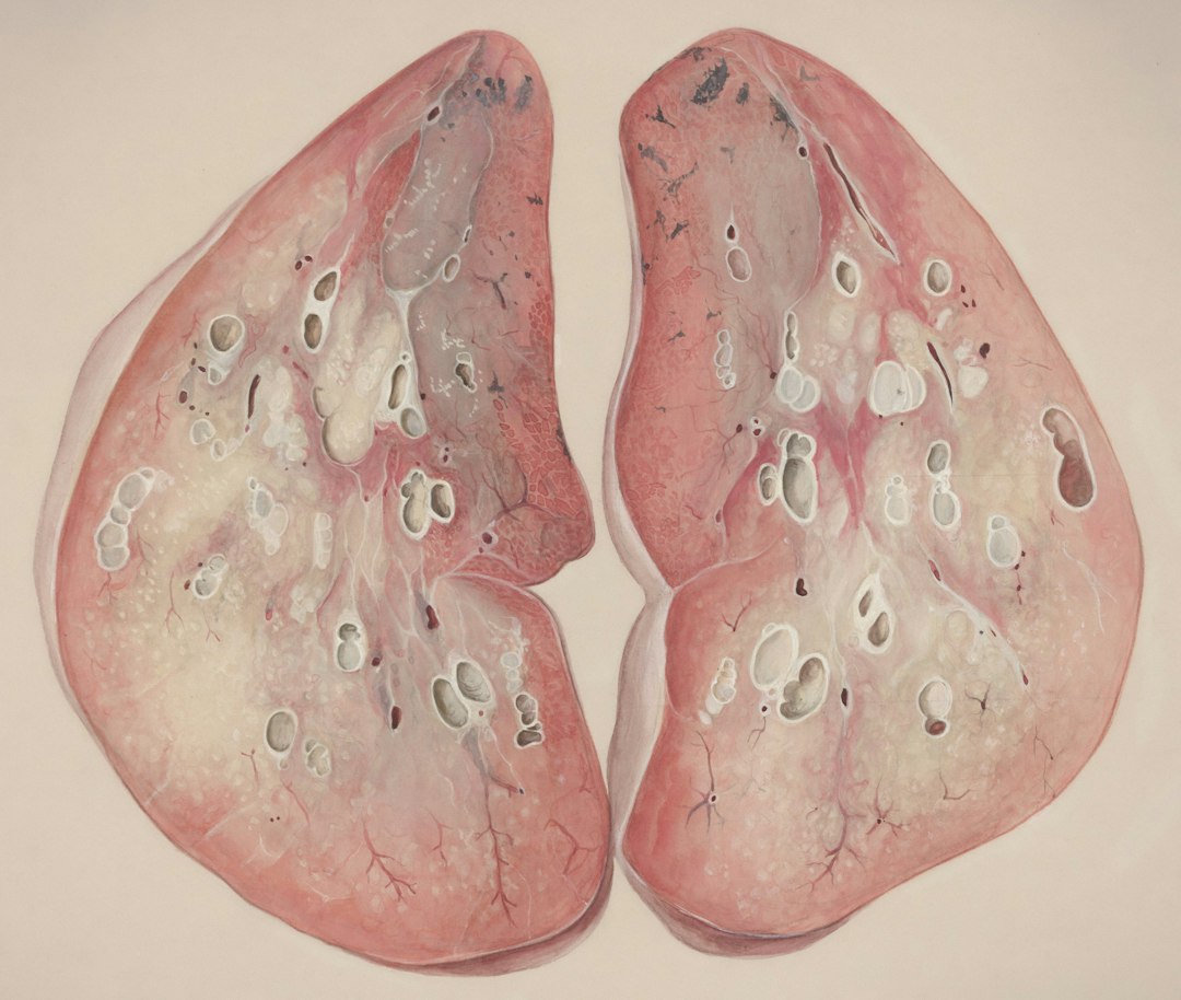


Title: Why Does It Hurt to Pee? A Complete Guide to Understanding, Treating, and Preventing Painful Urination

—

Introduction – The Uncomfortable “Why Me?” Moment

You’re in the middle of a busy day, you finally get a chance to use the restroom, and—ouch—a sharp sting hits you the moment the stream starts. The experience is not only painful; it’s also alarming, embarrassing, and often leaves you wondering, “What’s wrong with me?”

Painful urination, medically known as dysuria, is one of the most common urological complaints that brings people to the doctor’s office. While a single, mild episode may resolve on its own, recurring or severe pain can signal an infection, inflammation, or another underlying health issue that needs attention.

In this 2,000‑word deep dive we’ll explore:

  • What dysuria actually feels like and when it’s a red flag.
  • The top causes—from urinary tract infections (UTIs) to sexually transmitted infections (STIs).
  • How doctors diagnose the problem with simple, non‑invasive tests.
  • Proven treatment options, home remedies, and lifestyle tweaks that speed recovery.
  • Practical prevention strategies so you can keep the burn at bay.
  • Read on if you’ve ever felt that uncomfortable sting, or if you simply want to be equipped with the knowledge to protect yourself and your loved ones from painful urination.

    —

    1. What Is Painful Urination? Understanding Dysuria

    1.1 Defining Dysuria

    Dysuria is the medical term for pain, burning, or discomfort while urinating. The sensation can range from a mild irritation at the end of the stream to an intense, searing burn that makes it impossible to finish emptying the bladder. Dysuria can affect anyone, but it’s most common in women, children, and older adults.

    1.2 Typical Symptoms That Accompany Dysuria

    | Symptom | What It Means | Why It Matters |
    |———|—————|—————-|
    | Burning sensation during or after urination | Inflammation of the urethra or bladder lining | Often points to infection or irritation |
    | Frequent urge to urinate (urgency) | Bladder irritation | May indicate a UTI or interstitial cystitis |
    | Cloudy, foul‑smelling, or bloody urine | Presence of bacteria, blood, or pus | Signals infection or more serious pathology |
    | Pelvic or lower abdominal pain | Inflammation of surrounding organs | Can be a sign of kidney involvement |
    | Fever, chills, or flank pain | Systemic infection | Suggests kidney infection (pyelonephritis) |

    If you notice any combination of these signs—especially fever, flank pain, or blood in the urine—seek medical care promptly.

    1.3 When to Call a Doctor

    | Situation | Reason to Seek Immediate Care |
    |———–|——————————-|
    | Painful urination accompanied by fever > 100.4 °F (38 °C) | Possible kidney infection |
    | Blood in urine (hematuria) lasting more than a day | Could be infection, stones, or trauma |
    | Inability to urinate at all (urinary retention) | Emergency—risk of bladder damage |
    | Persistent dysuria for > 3 days without improvement | May need antibiotics or further work‑up |
    | Recent new sexual partner or unprotected sex + dysuria | Consider STI testing |

    —

    2. The Usual Suspects – Common Causes of Painful Urination

    Painful urination is a symptom, not a disease. Below are the most frequent culprits, broken down by category.

    2.1 Urinary Tract Infections (UTIs)

    UTIs are bacterial infections that can affect any part of the urinary system—urethra (urethritis), bladder (cystitis), ureters, or kidneys (pyelonephritis).

  • Why they cause dysuria: Bacteria invade the lining of the urinary tract, triggering inflammation and irritation.
  • Typical pathogens: Escherichia coli (E. coli) accounts for ~80 % of cases; other Gram‑negative rods, Enterococcus, and Klebsiella are also common.
  • Risk factors: Female anatomy (shorter urethra), sexual activity, pregnancy, catheter use, diabetes, and a history of prior UTIs.
  • 2.2 Sexually Transmitted Infections (STIs)

    STIs such as chlamydia, gonorrhea, and trichomoniasis can inflame the urethra (urethritis) and cause painful urination.

  • Key clues: Dysuria paired with genital discharge, itching, or sores.
  • Who’s at risk: Sexually active individuals, especially with multiple partners or inconsistent condom use.
  • 2.3 Bladder and Kidney Stones

    Hard mineral deposits can scrape the delicate lining of the urinary tract as they pass, resulting in a burning sensation.

  • Symptoms to watch: Sudden, severe flank pain that radiates to the groin, pink‑ish urine, and intermittent dysuria.
  • Common stones: Calcium oxalate, uric acid, struvite (often infection‑related).
  • 2.4 Prostatitis (Men)

    Inflammation of the prostate gland—either bacterial or non‑bacterial—can cause painful urination, pelvic pressure, and painful ejaculation.

  • Red flag: Chronic prostatitis may lead to persistent dysuria and reduced quality of life.
  • 2.5 Interstitial Cystitis (Painful Bladder Syndrome)

    A chronic condition where the bladder wall becomes inflamed without infection.

  • Typical presentation: Burning or pressure that worsens as the bladder fills, often accompanied by frequent urination (up to 60 times a day).
  • Triggers: Certain foods, stress, or unknown autoimmune factors.
  • 2.6 Vaginal Infections and Irritants (Women)

    Yeast infections, bacterial vaginosis, or even harsh soaps and douches can irritate the urethra, leading to dysuria.

  • Tip: If symptoms improve after stopping the irritant, you likely have a non‑infectious cause.
  • 2.7 Other Less Common Causes

    | Condition | How It Causes Dysuria |
    |———–|———————–|
    | Urinary catheter use | Direct irritation, biofilm formation |
    | Radiation therapy | Damage to bladder lining |
    | Neurological disorders (e.g., multiple sclerosis) | Disrupted nerve signals to bladder |
    | Medication side effects (e.g., cyclophosphamide) | Chemical irritation of urinary tract |

    —

    3. Getting to the Bottom of It – Diagnosis and Tests

    A proper diagnosis is the cornerstone of effective treatment. Most clinicians start with a focused history and a few simple tests.

    3.1 Medical History & Physical Exam

  • Symptom timeline: When did dysuria start? How severe? Any accompanying fever or flank pain?
  • Sexual history: Recent partners, condom use, known STI exposure.
  • Past urologic issues: Prior UTIs, kidney stones, surgeries.
  • Medication review: Any new drugs, especially antibiotics or chemotherapy agents.
  • A brief physical exam may include a pelvic exam (women) or digital rectal exam (men) to assess the prostate.

    3.2 Laboratory Tests

    | Test | What It Detects | Typical Result Interpretation |
    |——|—————–|——————————-|
    | Urinalysis (dip‑stick + microscopy) | Leukocytes, nitrites, blood, bacteria, crystals | Positive leukocyte esterase & nitrites → bacterial UTI |
    | Urine culture | Specific bacterial growth, antibiotic sensitivity | > 10⁵ CFU/mL of a single organism = infection |
    | STI panel (NAAT for chlamydia, gonorrhea, trichomonas) | Sexually transmitted pathogens | Positive → targeted antibiotic therapy |
    | Blood tests (CBC, CRP) | Systemic infection or inflammation | Elevated white blood cells → possible kidney infection |
    | Kidney function tests (creatinine, BUN) | Renal involvement | High levels may indicate pyelonephritis |

    3.3 Imaging & Specialized Studies

  • Ultrasound – Detects kidney stones, obstruction, or bladder wall thickening.
  • CT scan (non‑contrast) – Gold standard for identifying renal calculi.
  • Cystoscopy – Direct visual inspection of bladder and urethra, used for recurrent or unexplained dysuria.
  • Urodynamic testing – Assesses bladder function in cases of interstitial cystitis or neurogenic bladder.
  • 3.4 Putting It All Together

    A typical diagnostic pathway might look like this:

    1. Presenting complaint: Burning on urination for 2 days, no fever.
    2. Urinalysis: Positive leukocyte esterase, nitrites, and bacteria.
    3. Urine culture: Grows E. coli → sensitive to trimethoprim‑sulfamethoxazole.
    4. Diagnosis: Uncomplicated lower urinary tract infection (cystitis).
    5. Treatment: 3‑day course of antibiotics, increased fluid intake, and symptom relief measures.

    If the urinalysis is negative but dysuria persists, the clinician may pursue STI testing, imaging, or refer to a urologist for further evaluation.

    —

    4. Treatment Options – From Home Remedies to Prescription Therapy

    4.1 When Antibiotics Are the First Line

    For bacterial UTIs and many STIs, antibiotics remain the gold standard.

    | Condition | First‑line Antibiotic (Typical Regimen) |
    |———–|——————————————|
    | Uncomplicated cystitis (E. coli) | Trimethoprim‑sulfamethoxazole 160/800 mg BID for 3 days |
    | Complicated UTI or pyelonephritis | Ciprofloxacin 500 mg BID for 7–14 days |
    | Chlamydia urethritis | Azithromycin 1 g PO single dose |
    | Gonorrhea urethritis | Ceftriaxone 500 mg IM single dose + doxycycline 100 mg BID for 7 days (if chlamydia co‑infection suspected) |

    Key tip: Always complete the full antibiotic course, even if symptoms improve within 48 hours, to prevent resistance and relapse.

    4.2 Pain Relief and Symptom Management

  • Phenazopyridine (e.g., Pyridium) – OTC urinary analgesic that provides temporary relief of burning and urgency. Use for no more than 2 days to avoid masking worsening infection.
  • NSAIDs (ibuprofen, naproxen) – Reduce inflammation and lower fever.
  • Heat therapy – Warm compresses on the lower abdomen can soothe muscle spasms.
  • 4.3 Home Care Strategies

    | Action | How It Helps |
    |——–|————–|
    | Increase fluid intake (aim for 2–3 L water/day) | Flushes bacteria from the bladder; dilutes urine, reducing irritation |
    | Urinate frequently (every 2–3 hours) | Prevents bacterial overgrowth and reduces bladder pressure |
    | Cranberry products (unsweetened juice or capsules) | May prevent bacterial adhesion to bladder walls (evidence mixed; best as adjunct) |
    | Avoid bladder irritants (caffeine, alcohol, spicy foods, artificial sweeteners) | Reduces inflammation and urgency |
    | Proper hygiene – front‑to‑back wiping, urinating after intercourse | Lowers bacterial transfer to urethra |
    | Probiotic yogurt or supplements | Supports healthy vaginal flora, especially after antibiotics |

    4.4 Managing Non‑Bacterial Causes

  • Interstitial cystitis: Oral pentosan polysulfate, bladder instillations, pelvic floor physical therapy, and dietary modifications (low‑acid, low‑caffeine diet).
  • Prostatitis: Alpha‑blockers (tamsulosin) for symptom relief, plus a 2–4‑week course of appropriate antibiotics if bacterial.
  • Kidney stones: Hydration, pain control, and, when needed, lithotripsy or surgical removal.
  • Vaginal infections: Antifungal (fluconazole) for yeast, metronidazole for bacterial vaginosis, and avoidance of scented hygiene products.
  • 4.5 Follow‑Up and When to Re‑Evaluate

  • UTI: Re‑check urine culture if symptoms persist after 48–72 hours of antibiotics.
  • STI: Re‑test in 3 months (or sooner if re‑exposure) to confirm cure.
  • Recurrent dysuria (≥ 3 episodes per year): Consider a urology referral for cystoscopy or further work

Title: Dialysis Demystified: A Complete Guide to Kidney Replacement Therapy, What to Expect, and How to Thrive

—

Introduction – Why “Dialysis” Is More Than a Medical Term

Imagine your kidneys as a sophisticated filtration plant that works 24/7, cleaning blood, balancing electrolytes, and removing waste. When that plant falters, the body’s internal chemistry spirals out of control—fatigue, swelling, nausea, and life‑threatening complications can follow. For millions worldwide, dialysis is the lifeline that steps in when kidneys can no longer do their job.

But “dialysis” often feels like a mysterious, intimidating word whispered in hospital corridors. Is it a one‑time procedure? A lifelong commitment? Which type is right for you or a loved one? How do diet, lifestyle, and mental health fit into the picture?

This comprehensive, 2,000‑word guide pulls back the curtain on renal replacement therapy. We’ll break down the science, compare the main dialysis modalities, walk you through the practical steps of getting started, and share actionable tips to help patients and caregivers not just survive—but thrive—on dialysis. Whether you’re a newly diagnosed chronic kidney disease (CKD) patient, a family member seeking clarity, or a health‑conscious reader curious about the topic, you’ll finish this article with confidence and a clear roadmap.

—

1. Understanding Dialysis: The “Why” and the “How”

1.1 What Exactly Is Dialysis?

Dialysis is an artificial process that mimics the kidney’s natural filtering function. By moving blood through a semi‑permeable membrane, dialysis removes excess fluid, urea, electrolytes (like potassium and phosphorus), and metabolic waste, while returning essential substances back to the bloodstream.

Key terms you’ll hear:

  • Renal replacement therapy (RRT): The umbrella term for dialysis and kidney transplantation.
  • Uremia: The buildup of waste products in the blood that dialysis aims to clear.
  • Glomerular filtration rate (GFR): A measure of kidney function; dialysis is typically recommended when GFR falls below 15 mL/min/1.73 m².
  • 1.2 When Is Dialysis Needed?

    Dialysis isn’t prescribed solely based on a lab number. Clinicians weigh a combination of factors:

    | Clinical Indicator | Typical Threshold | Why It Matters |
    |——————–|——————-|—————-|
    | GFR | < 15 mL/min/1.73 m² (Stage 5 CKD) | Indicates severe loss of filtering capacity |
    | Fluid overload | Persistent edema, shortness of breath | Excess fluid can cause heart failure |
    | Electrolyte imbalance | Hyperkalemia (K⁺ > 6.0 mmol/L) | Dangerous for cardiac rhythm |
    | Uremic symptoms | Nausea, itching, pericarditis, mental status changes | Signifies toxin buildup |
    | Acidosis | Bicarbonate < 18 mmol/L | Leads to bone disease & muscle wasting |

    If any of these red flags appear, the nephrologist will discuss dialysis options, often alongside transplant evaluation.

    1.3 The Two Main Types of Dialysis

    | Modality | How It Works | Typical Frequency | Pros | Cons |
    |———-|————–|——————-|——|——|
    | Hemodialysis (HD) | Blood pumped through an external machine; waste diffuses across a dialyzer (artificial kidney). | 3‑times weekly, ~4 hours per session (in‑center) or 5‑6 times weekly (home). | Rapid clearance; close monitoring by staff; no need for daily self‑care. | Travel to center; vascular access surgery; “dialysis fatigue” after sessions. |
    | Peritoneal Dialysis (PD) | Fluid infused into the abdomen; peritoneal membrane acts as filter; waste moves into fluid, which is drained. | 4‑5 exchanges daily (continuous ambulatory PD) or automated overnight (APD). | Greater flexibility; can be done at home; gentler fluid shifts. | Requires catheter care; risk of peritonitis; storage space for supplies. |

    Both modalities are considered renal replacement therapy and can be life‑saving. The choice hinges on medical suitability, lifestyle preferences, support system, and personal values.

    —

    2. Getting Started: Preparing for Dialysis Success

    2.1 Vascular Access – The Gateway to Effective Treatment

    For Hemodialysis:

  • Arteriovenous fistula (AVF): Surgical connection of an artery to a vein; gold standard because it lasts years and has low infection risk.
  • Arteriovenous graft (AVG): Synthetic tube linking artery and vein; used when veins are too small for a fistula.
  • Central venous catheter (CVC): Temporary line placed in a large vein; higher infection risk; used only while waiting for a fistula/graft to mature.
  • For Peritoneal Dialysis:

  • Tenckhoff catheter: Soft, flexible tube placed into the peritoneal cavity under local anesthesia; must heal for ~2 weeks before first exchange.
  • Actionable tip:
    Schedule your access surgery early. AVFs take 4‑8 weeks to mature. Early placement gives you time for healing, reduces reliance on catheters, and improves long‑term outcomes.

    2.2 The First Dialysis Session – What to Expect

    | Step | What Happens | How to Prepare |
    |——|————–|—————-|
    | Pre‑session labs | Blood work (CBC, electrolytes, calcium/phosphate) to fine‑tune the prescription. | Fast as instructed (usually 8 hrs). Bring a list of meds. |
    | Access check | Nurse verifies fistula “thrill” or catheter patency. | Keep the access site clean; avoid tight clothing. |
    | Machine set‑up | Dialyzer, tubing, and dialysate are prepared. | Arrive early; bring a water bottle, headphones, or a book. |
    | Treatment | Blood flows at 300‑500 mL/min; waste diffuses into dialysate. | Stay still, relax, and use the time for light activity (e.g., knitting). |
    | Post‑treatment | Blood pressure check, weight measurement, and access inspection. | Record your post‑dialysis weight; note any cramps or dizziness. |

    Most patients feel a “dialysis hangover” after the first few sessions—light fatigue, mild nausea, or muscle cramps. This usually improves as your body adapts.

    2.3 Building a Support Network

    Dialysis can be overwhelming, but you don’t have to navigate it alone. Here’s a quick checklist:

  • Nephrology team: Doctor, nurse, dietitian, social worker.
  • Family & friends: Identify a “dialysis buddy” for rides or catheter care.
  • Patient groups: Local kidney foundations or online forums (e.g., National Kidney Foundation, Renal Support Network).
  • Mental health resources: Counseling or mindfulness apps to manage anxiety.
  • Pro tip: Keep a “dialysis diary” (paper or digital) to track symptoms, fluid intake, weight, and mood. Share it with your care team at each visit.

    —

    3. Living the Dialysis Lifestyle – Nutrition, Exercise, and Daily Routines

    3.1 Dialysis‑Friendly Diet: Balancing Fluids, Sodium, Potassium, and Protein

    Dialysis changes how your body handles nutrients. Below is a practical, food‑focused guide.

    | Nutrient | Why It Matters | Recommended Target (average adult) | Easy Food Swaps |
    |———-|—————-|———————————–|—————–|
    | Protein | Helps rebuild tissues; dialysis removes some protein. | 1.2‑1.4 g/kg body weight per day (HD); 1.0‑1.2 g/kg (PD). | Choose lean poultry, fish, eggs, tofu; limit processed meats. |
    | Sodium | Controls fluid retention and blood pressure. | < 2,300 mg/day (≈1 tsp salt). | Use herbs, lemon, garlic instead of table salt. |
    | Potassium | Prevents dangerous heart rhythm changes. | 2,000‑3,000 mg/day (depends on labs). | Swap banana for apples; use cauliflower rice instead of potatoes. |
    | Phosphorus | Protects bones and blood vessels. | 800‑1,000 mg/day. | Choose fresh meats over cheese; avoid cola and processed cheese. |
    | Fluids | Avoids overload that stresses the heart. | Typically 1‑1.5 L per day for HD; may be higher for PD (depends on residual kidney function). | Measure all drinks; use a marked water bottle. |

    Actionable daily plan (example for a 70 kg HD patient):

  • Breakfast: Scrambled egg whites + spinach + slice whole‑grain toast; 1 cup orange juice (low‑potassium).
  • Mid‑morning snack: Small apple + 10 g unsalted almonds.
  • Lunch: Grilled chicken breast, quinoa, steamed green beans, salad with olive oil vinaigrette; water (200 ml).
  • Afternoon snack: Rice cakes with low‑phosphate cream cheese.
  • Dinner: Baked cod, mashed cauliflower, sautéed zucchini; water (200 ml).
  • Evening: Herbal tea (no added sugar) + 1 small piece of low‑phosphate fruit (e.g., berries).
  • Always discuss your individualized targets with a renal dietitian—lab values will guide adjustments.

    3.2 Exercise on Dialysis: Keeping the Heart Strong

    Physical activity improves cardiovascular health, mood, and dialysis efficiency. Here’s a safe, step‑by‑step approach:

    1. Start Slow: 10‑15 minutes of walking or stationary cycling on non‑dialysis days.
    2. In‑Session Exercise: Light resistance bands or hand‑grip squeezers during HD (if the nurse approves).
    Benefit: Reduces muscle cramps and improves blood flow.
    3. Strength Training: 2‑3 sessions per week of body‑weight exercises (squats, wall push‑ups).
    Tip: Use a chair for support if balance is an issue.
    4. Flexibility & Balance: Yoga or Tai Chi once a week to enhance joint mobility and reduce fall risk.

    Safety check: Check blood pressure and blood sugar (if diabetic) before and after exercise. If you feel dizzy, short‑of‑breath, or experience chest pain, stop and call your care team.

    3.3 Managing Medications and Common Symptoms

    Dialysis changes how drugs are cleared, so medication management is critical.

    | Symptom | Typical Medication | Dialysis Considerations |
    |———|——————–|————————–|
    | Anemia | Erythropoiesis‑stimulating agents (ESA) + iron supplements | ESA dose adjusted based on hemoglobin; IV iron given during HD. |
    | Bone‑mineral disorder | Phosphate binders (sevelamer, calcium acetate) | Take with meals; avoid chewable binders if you have swallowing issues. |
    | High blood pressure | ACE inhibitors, ARBs, beta‑blockers | Some are removed during HD; dosing may need timing around sessions. |
    | Pruritus (itching) | Antihistamines, gabapentin | Gabapentin dose reduced for HD patients. |
    | Nausea | Ondansetron, prochlorperazine | Generally safe; monitor for QT prolongation. |

    Practical tip: Keep an up‑to‑date medication list on your fridge. Mark which pills are taken on dialysis days versus non‑dialysis days.

    —

    4. Choosing Between Hemodialysis and Peritoneal Dialysis – A Decision‑Making Toolkit

    4.1 Lifestyle Compatibility Checklist

    | Factor | Hemodialysis (In‑Center) | Hemodialysis (Home) | Peritoneal Dialysis |
    |——–|————————–|———————|———————|
    | Work schedule | Fixed 3×/week visits; may need time off | Flexible if you own a machine | Can be done overnight (APD) or daytime (CAPD) |
    | Travel | Requires proximity to a dialysis unit | Portable machines available | Supplies can be mailed; need clean space |
    | Home environment | Not required | Requires dedicated space for machine and water treatment | Needs clean, dry area for supplies and a place for exchanges |
    | Support system | Staff handles most tasks | You or a caregiver manage setup | You or a caregiver must perform exchanges |
    | Medical suitability | Good vascular access | Same as in‑center HD | Intact peritoneal membrane; no severe abdominal adhesions |
    | Preference for independence | Lower | Higher | Highest (especially APD) |

    Self‑assessment: Score each factor from 1 (low priority) to 5 (high priority). Add the scores for each modality; the highest total suggests the best fit, but always discuss with your nephrologist.

    4.2 Cost & Insurance Considerations

  • In‑center HD: Typically covered fully by Medicare, Medicaid, and most private insurers in the U.S., but indirect costs (travel, parking, time off work) add up.
  • Home HD: Higher upfront equipment cost; many insurers provide a “

Title: Prostate Cancer Uncovered: Early Signs, Diagnosis, Treatment & Living Well Beyond the Diagnosis

—

Introduction – Why This Talk About Prostate Cancer Matters Now

Imagine you’re scrolling through your phone, reading the latest headlines, when a statistic pops up: 1 in 8 men will be diagnosed with prostate cancer in their lifetime. For many, that number feels abstract—until it hits close to home. Whether you’re a man approaching middle age, a partner, a caregiver, or simply a health‑conscious individual, understanding prostate cancer is no longer optional; it’s essential.

In this comprehensive guide we’ll break down the most common questions about prostate cancer, from the subtle early signs you might overlook to the latest breakthroughs in treatment. You’ll walk away with actionable steps you can take today—whether that means scheduling a PSA test, tweaking your diet, or navigating survivorship after treatment. Let’s demystify the disease, empower you with knowledge, and show that early detection truly saves lives.

—

1. Understanding Prostate Cancer: The Basics You Need to Know

What Is the Prostate and Why Does It Matter?

The prostate is a walnut‑sized gland located just below the bladder, surrounding the urethra—the tube that carries urine (and semen) out of the body. Its primary job is to produce fluid that nourishes and protects sperm. Because of its location, any abnormal growth can affect urinary function, sexual health, and overall well‑being.

How Common Is Prostate Cancer?

  • Incidence: Over 250,000 new cases are diagnosed in the United States each year.
  • Mortality: It’s the second leading cause of cancer death among men, but the 5‑year survival rate exceeds 95% when caught early.
  • Age Factor: The risk climbs sharply after age 50; about 60% of diagnoses occur after 65.
  • Key Risk Factors (And What You Can Do About Them)

    | Risk Factor | Why It Increases Risk | Actionable Tips |
    |————-|———————-|—————–|
    | Age | Cellular changes accumulate over time. | Begin regular screenings at 45 (or 40 if you have a family history). |
    | Family History | Genetics can predispose you to aggressive forms. | Discuss your pedigree with a doctor; consider earlier PSA testing. |
    | African‑American Heritage | Higher incidence and mortality rates. | Prioritize early screening; maintain a heart‑healthy lifestyle. |
    | Diet & Lifestyle | High‑fat, low‑fiber diets linked to inflammation. | Adopt a Mediterranean‑style diet rich in tomatoes, fish, and whole grains. |
    | Obesity | Hormonal imbalances may fuel tumor growth. | Aim for a BMI < 25; incorporate regular cardio and strength training. |
    | Exposure to Certain Chemicals (e.g., Agent Orange) | Disrupts hormonal pathways. | Limit exposure; discuss occupational risks with your physician. |

    > Pro Tip: Even if you lack obvious risk factors, age alone justifies regular check‑ups. Prostate cancer often progresses silently, so proactive monitoring is your best defense.

    The Biology in Plain English

    Prostate cancer starts when a single prostate cell mutates, begins dividing uncontrollably, and forms a tumor. Most tumors are adenocarcinomas—cancers arising from glandular tissue. They can be:

  • Low‑grade (Gleason score ≤ 6) – slow‑growing, often managed with active surveillance.
  • Intermediate‑grade (Gleason 7) – may need a combination of surgery, radiation, or hormone therapy.
  • High‑grade (Gleason 8‑10) – aggressive, requiring multimodal treatment.
  • Understanding the Gleason score and staging (TNM system) helps you and your doctor decide the most appropriate treatment plan.

    —

    2. Spotting the Early Warning Signs – When to Get Checked

    Prostate cancer is notorious for being “silent” in its early stages. However, a few subtle cues can signal that something’s amiss. Keep an eye out for the following symptoms and red flags:

    | Symptom | What It Might Indicate | When to Seek Medical Advice |
    |———|———————–|—————————–|
    | Frequent urination, especially at night | Enlarged prostate or early tumor | If >2–3 times nightly for >1 month |
    | Weak or interrupted urine stream | Possible obstruction | Persistent changes >2 weeks |
    | Blood in urine or semen | Possible tumor bleeding | Any visible blood warrants prompt evaluation |
    | Painful ejaculation | Irritation or tumor involvement | Ongoing discomfort |
    | Persistent pelvic or lower back pain | Advanced disease spreading to bones | Pain lasting >2 weeks |
    | Unexplained weight loss | Systemic effect of cancer | Any rapid loss >5% body weight |

    > Action Step: Keep a simple symptom diary. Note frequency, intensity, and any triggers. Bring this log to your next primary‑care visit—doctors love concrete data.

    PSA Test: The Screening Cornerstone

    The Prostate‑Specific Antigen (PSA) blood test measures a protein produced by both normal and malignant prostate cells. While PSA isn’t a perfect marker (it can rise due to inflammation or benign enlargement), it remains the most widely used screening tool.

    How to Interpret PSA Results

    | PSA Level (ng/mL) | Typical Interpretation |
    |——————-|————————|
    | < 4.0 | Generally low risk, but not zero |
    | 4.0 – 10.0 | “Gray zone”; consider repeat testing, free/total PSA ratio, or MRI |
    | > 10.0 | Higher probability of cancer; further diagnostic work‑up needed |

    Tips for Optimizing PSA Accuracy

    1. Avoid PSA‑affecting activities (e.g., vigorous bike riding, recent ejaculation) 48‑72 hours before the test.
    2. Discuss medications (like 5‑alpha‑reducing drugs) that can lower PSA and mask disease.
    3. Ask about PSA velocity (rate of change over time) – a rapid rise may be more concerning than a single high value.

    —

    3. Diagnosis Pathway – From Suspicion to Confirmation

    If your PSA or digital rectal exam (DRE) raises concerns, your doctor will guide you through a step‑by‑step diagnostic process.

    3.1 Digital Rectal Exam (DRE)

    A quick, in‑office exam where the physician feels the prostate through the rectal wall to detect lumps, hard areas, or asymmetry. While not definitive, an abnormal DRE often prompts further testing.

    3.2 Imaging: Multiparametric MRI (mpMRI)

    Modern guidelines increasingly recommend a multiparametric MRI before a biopsy. mpMRI can:

  • Detect clinically significant tumors.
  • Reduce unnecessary biopsies.
  • Help target biopsy needles to suspicious areas (fusion‑guided biopsy).
  • What to Expect: The scan takes ~30‑45 minutes, no contrast is required for most protocols, and you’ll lie still in a tube-like machine.

    3.3 Prostate Biopsy – The Definitive Test

    If imaging suggests cancer, a prostate biopsy is performed—usually via a transrectal ultrasound (TRUS) or transperineal approach. The doctor extracts 10‑12 core tissue samples for pathological analysis.

    Emerging Alternatives

  • MRI‑targeted biopsy (fewer cores, higher detection of aggressive disease).
  • Liquid biopsy (blood test for circulating tumor DNA) – still investigational but promising for future monitoring.
  • 3.4 Staging – How Far Has It Spread?

    Once cancer is confirmed, staging determines the extent:

  • TNM System (Tumor size, Node involvement, Metastasis).
  • Gleason Score – grades tumor architecture.
  • PSA Level – helps predict tumor burden.
  • Staging Tools

  • Bone Scan – detects skeletal metastases (common in advanced disease).
  • CT or PET Scan – evaluates lymph nodes and distant organs.
  • > Bottom Line: Accurate staging guides treatment choices and prognosis. Ask your oncologist to explain each result in plain language.

    —

    4. Treatment Options – Tailoring Care to Your Situation

    Prostate cancer treatment isn’t one‑size‑fits‑all. Choices depend on age, overall health, cancer stage, Gleason score, and personal preferences. Below is a concise roadmap of standard and emerging therapies, along with actionable considerations for each.

    4.1 Active Surveillance (Watchful Waiting)

  • Who It’s For: Low‑grade (Gleason ≤ 6), localized tumors, typically men >65 with limited life expectancy or those preferring to avoid side effects.
  • What It Involves: Regular PSA testing every 3‑6 months, annual MRI, and repeat biopsies every 1‑3 years.
  • Pros: Avoids or delays surgery/radiation; preserves urinary and sexual function.
  • Cons: Requires strict adherence to follow‑up; anxiety for some patients.
  • Actionable Tip: If you choose active surveillance, set calendar reminders for each PSA check and keep a log of results. Share this log with your urologist at each visit.

    4.2 Surgery – Radical Prostatectomy

  • Procedure: Removal of the entire prostate gland and surrounding tissue, usually via open, laparoscopic, or robot‑assisted (da Vinci) approaches.
  • Ideal Candidates: Men with localized disease (stage T1‑T2), good overall health, and a desire for definitive treatment.
  • Potential Side Effects: Urinary incontinence, erectile dysfunction, and rare bowel complications.
  • Recovery Hacks

    1. Pelvic floor exercises (Kegels) start within days post‑op to speed continence recovery.
    2. Warm compresses reduce swelling and promote healing.
    3. Nutrition: High‑protein, low‑sugar meals aid tissue repair.

    4.3 Radiation Therapy

  • External Beam Radiation Therapy (EBRT): Daily high‑energy X‑rays over 6‑8 weeks.
  • Brachytherapy (Seed Implant): Radioactive seeds placed directly into the prostate, delivering localized dose over months.
  • Combination (EBRT + Brachy): For intermediate‑high risk disease.
  • Side Effects & Management

    | Side Effect | Typical Onset | Management Strategies |
    |————-|—————|————————|
    | Fatigue | Weeks 2‑4 | Light exercise, sleep hygiene |
    | Urinary urgency | First few weeks | Alpha‑blockers (e.g., tamsulosin) |
    | Rectal irritation | Ongoing | Dietary fiber, stool softeners |
    | Erectile dysfunction | 3‑6 months | PDE5 inhibitors, counseling |

    4.4 Hormone Therapy (Androgen Deprivation Therapy – ADT)

    Prostate cancer cells thrive on testosterone. ADT reduces androgen levels using:

  • LHRH agonists/antagonists (e.g., leuprolide, degarelix).
  • Anti‑androgens (e.g., bicalutamide, enzalutamide).
  • When Used: Often combined with radiation for high‑risk disease or as a bridge to other treatments in metastatic settings.

    Side Effect Checklist

  • Hot flashes
  • Bone density loss → Action: Start weight‑bearing exercise + calcium/vitamin D; consider a DEXA scan.
  • Metabolic changes → Monitor blood sugar and cholesterol.
  • 4.5 Chemotherapy & Targeted Therapy

  • Docetaxel is the standard chemo for metastatic castration‑resistant prostate cancer (mCRPC).
  • PARP inhibitors (e.g., olaparib) for tumors with DNA‑repair gene mutations (BRCA1/2).
  • Immunotherapy (Sipuleucel‑T) for select mCRPC patients.

Key Consideration: Discuss clinical trial eligibility. Many cutting‑edge therapies are only available through research protocols.

4.6 Emerging Treatments & Lifestyle Adjuncts

| Innovation | Current Status | Practical Takeaway |
|————|—————-|——————–|
| PSMA‑targeted radioligand therapy (e.g., Lutetium‑177‑PSMA) | FDA‑approved for mCRPC (2022) | Offers targeted radiation with fewer systemic side effects. |
| High‑Intensity Focused Ultrasound (HIFU) | Investigational for localized disease | Non‑invasive, outpatient; may preserve function better. |
| Nutraceuticals (e.g., lycopene, green tea catechins) | Mixed evidence | Incorporate tomato‑based sauces and green tea; no substitute for medical therapy. |

> Action Plan: Ask your oncologist about clinical trials in your area. Even if you’re not eligible now, staying informed keeps you at the forefront of care.

—

5. Living Beyond Diagnosis – Survivorship, Mental Health, and Prevention

A prostate cancer diagnosis can feel like a life‑changing event, but many men go on to lead vibrant, fulfilling lives. The survivorship phase focuses on quality of life, long‑term monitoring, and preventive health.

5.1 Follow‑Up Schedule

| Time Post‑Treatment | Recommended Tests |
|———————|——————–|
| 3‑6 months | PSA, physical exam |
| Every 6 months (

Title: Strengthen, Support, and Thrive: The Ultimate Guide to Pelvic Floor Exercises

—

Introduction – Why the Pelvic Floor Deserves Your Attention

Imagine a hammock that holds up every organ in your lower abdomen, supports your spine, and even helps you control the most private moments of your life. That “hammock” is your pelvic floor—a deep, circular set of muscles that many people never think about—until something goes wrong. From urinary leaks during a laugh, to low‑back pain that won’t quit, to a sluggish recovery after childbirth, a weak pelvic floor can silently sabotage your confidence and comfort.

The good news? You can train these muscles just like you would your biceps or glutes, and you don’t need fancy equipment or a gym membership. With consistent pelvic floor exercises—commonly known as Kegels, but far more varied—you can boost core stability, improve sexual health, and reclaim control over your body. This comprehensive guide walks you through the science, the step‑by‑step techniques, and the everyday habits that turn a neglected muscle group into a powerhouse of support. Let’s dive in and give your pelvic floor the attention it deserves.

—

1. Understanding the Pelvic Floor: Anatomy, Functions, and Common Issues

1.1 What Is the Pelvic Floor?

The pelvic floor is a sling of muscles, ligaments, and fascia that stretches like a bowl from the pubic bone in front to the tailbone at the back. It forms the base of the core, cradling the bladder, uterus (or prostate), and rectum. Key muscle groups include the pubococcygeus (PC), iliococcygeus, and puborectalis—collectively known as the levator ani.

1.2 Why It Matters

  • Urinary and bowel control – The pelvic floor contracts to close the urethra and anus, preventing leaks.
  • Core stability – It works with the transverse abdominis and diaphragm to stabilize the spine during movement.
  • Sexual function – Strong pelvic muscles improve blood flow and sensation, enhancing orgasmic potential.
  • Postpartum recovery – After pregnancy, the floor stretches dramatically; rebuilding strength helps prevent prolapse and incontinence.
  • 1.3 Common Pelvic Floor Problems

    | Issue | Typical Symptoms | Who’s at Risk |
    |——-|——————|—————-|
    | Stress urinary incontinence | Leakage when coughing, sneezing, or exercising | Women post‑childbirth, older adults |
    | Pelvic organ prolapse | A feeling of heaviness, bulge in the vagina or rectum | Women with multiple vaginal deliveries |
    | Chronic pelvic pain | Persistent ache in the lower abdomen/pelvis | Anyone with muscle tension or nerve irritation |
    | Erectile dysfunction (men) | Difficulty achieving/maintaining erection | Men with weak pelvic muscles or after prostate surgery |

    Understanding the root cause helps you pick the right exercises and avoid “one‑size‑fits‑all” routines that may aggravate the problem.

    —

    2. Mastering the Basics: Kegel Exercises and Their Variations

    2.1 The Classic Kegel (Step‑by‑Step)

    1. Locate the muscles – Imagine trying to stop the flow of urine mid‑stream. The muscles you contract are your pelvic floor.
    2. Get comfortable – Sit or lie down with knees bent, feet flat. Relax the abdomen, buttocks, and thighs.
    3. Contract – Squeeze the pelvic floor for 3–5 seconds, then fully relax for the same amount of time.
    4. Repeat – Aim for 10 repetitions, three times a day.

    Pro tip: Use a timer or a smartphone app that cues “inhale, hold, exhale” to keep the rhythm consistent.

    2.2 Advanced Kegel Variations

    | Variation | How to Do It | Benefits |
    |———–|————–|———-|
    | Quick flicks | Contract and release the pelvic floor as fast as possible for 10–15 reps. | Improves fast‑twitch muscle fibers, helpful for sudden stress leaks. |
    | Elevator lifts | Imagine your pelvic floor as an elevator: start with a gentle squeeze, then gradually increase the intensity in three “floors,” hold at the top, then descend. | Builds endurance and teaches gradated control. |
    | Supine hip thrust Kegels | Lie on your back, knees bent, lift hips slightly off the floor, then perform a Kegel while the glutes are engaged. | Integrates pelvic floor with glute activation for functional core stability. |
    | Standing pelvic squeezes | While standing, gently draw the pelvic floor upward as if “pulling your belly button toward your spine.” | Trains the muscles in a functional, weight‑bearing position. |

    2.3 Common Mistakes and How to Fix Them

  • Holding the breath – The diaphragm should stay relaxed; breathe naturally.
  • Engaging the glutes or abs – Focus on a subtle lift under the perineum, not a full buttock squeeze.
  • Over‑contracting – Too much pressure can cause pelvic pain; aim for a gentle, controlled contraction.
  • If you’re unsure whether you’re doing it right, try the “stop‑the‑stream” test: interrupt your urine flow once, then finish the stream. That brief interruption confirms you’ve isolated the correct muscles. (Don’t make a habit of stopping mid‑stream—just use it as a learning tool.)

    —

    3. Integrating Pelvic Floor Work Into Your Daily Routine

    3.1 Building a Consistent Schedule

    | Time of Day | Activity | How to Incorporate |
    |————-|———-|——————–|
    | Morning | Bathroom routine | Perform a set of 5 quick flicks while brushing teeth. |
    | Mid‑day | Desk work | Do “standing pelvic squeezes” during a coffee break (10 reps). |
    | Evening | TV time | While watching your favorite show, complete a full Kegel set (10 reps). |
    | Bedtime | Wind‑down | Finish the day with an “elevator lift” series, focusing on slow, deep holds. |

    Consistency beats intensity. Even five seconds of a proper contraction a few times a day adds up over weeks.

    3.2 Pairing Pelvic Floor Exercises With Core Work

    Your pelvic floor is a teammate of the transverse abdominis (TA), the deepest abdominal muscle. Strengthening them together creates a “core corset” that protects the spine.

  • Dead Bug with Kegels – Lie on your back, arms toward the ceiling, knees bent at 90°. As you extend the right arm and left leg, engage your pelvic floor and TA. Return to start, then switch sides.
  • Bird‑Dog + Pelvic Lift – From all fours, extend opposite arm and leg while gently pulling the pelvic floor upward. This trains coordination and stability.
  • 3.3 Lifestyle Hacks for Pelvic Health

    1. Stay hydrated – Adequate fluid intake prevents bladder irritation, making training easier.
    2. Fiber‑rich diet – Prevent constipation, which can strain the pelvic floor during bowel movements.
    3. Mindful posture – Slouching compresses the pelvis; sit tall, engage the core, and keep the pelvis in a neutral tilt.
    4. Avoid heavy lifting without bracing – When you lift, inhale, brace the core (including the pelvic floor), and exhale after the lift.

    —

    4. Special Populations: Postpartum, Men, and Seniors

    4.1 Postpartum Recovery – Rebuilding After Birth

    Childbirth stretches the pelvic floor dramatically, especially after a vaginal delivery. Here’s a safe progression:

    | Week Post‑Birth | Focus | Sample Exercise |
    |—————-|——-|—————–|
    | 0–2 | Gentle activation | Pelvic tilts: Lie on your back, knees bent, gently flatten the lower back while subtly pulling the pelvic floor up. |
    | 3–6 | Endurance | Supine Kegels: 10‑second holds, 8 reps, twice daily. |
    | 7–12 | Functional strength | Squat to chair with Kegels: Perform a shallow squat, engage the pelvic floor on the way up. |
    | 13+ | Return to sport | Lunge with pelvic lift: Add a Kegel to each lunge, progressing to weighted lunges if cleared by a PT. |

    Always get clearance from your OB‑GYN or a pelvic health physiotherapist before starting a vigorous program.

    4.2 Men’s Pelvic Floor Health

    Men often overlook pelvic health until they experience prostate surgery or erectile dysfunction. The same principles apply:

  • Kegel for men – Contract the muscles you’d use to stop passing gas.
  • Reverse Kegels – Gently relax the floor to improve pelvic blood flow and reduce tension.
  • Hip thrusts – Strengthen glutes and the posterior chain, which support pelvic alignment.
  • A study in The Journal of Urology (2022) showed that a 12‑week pelvic floor program reduced post‑prostatectomy urinary leakage by 45 %.

    4.3 Seniors – Maintaining Independence

    Aging brings natural muscle atrophy, but targeted pelvic floor work can preserve continence and balance:

  • Seated Kegels – Ideal for those with limited mobility.
  • Chair yoga with pelvic focus – Poses like Seated Cat‑Cow encourage diaphragmatic breathing and gentle pelvic engagement.
  • Resistance band “hip abduction + Kegel” – While seated, press a mini‑band around the thighs, abduct the legs, and contract the pelvic floor simultaneously.
  • Regular pelvic floor training has been linked to a 30 % lower risk of falls in women over 65, thanks to improved core stability.

    —

    5. Tracking Progress and Knowing When to Seek Professional Help

    5.1 Simple Self‑Assessment Tools

  • The “Stop‑the‑Stream” Test – As mentioned, a quick way to confirm you’re using the right muscles.
  • Pelvic Floor Muscle Strength Scale (Oxford Scale) – Ranges from 0 (no contraction) to 5 (strong contraction). Rate yourself after a few weeks of practice; aim for a 3 or higher.
  • Leak Diary – Record any episodes of urinary leakage, activity triggers, and whether you performed a Kegel before the event. Patterns reveal improvement over time.
  • 5.2 When to Call a Pelvic Health Professional

  • Persistent leakage despite 4–6 weeks of consistent training.
  • Pain during Kegels or a feeling of “tightness” that doesn’t relax.
  • Noticeable bulge or pressure in the vaginal or rectal area (possible prolapse).
  • Post‑surgical concerns (e.g., after hysterectomy or prostatectomy).
  • A pelvic floor physical therapist can perform a digital exam, biofeedback, or electrical stimulation to fine‑tune your program.

    5.3 Tech‑Assisted Training

  • Biofeedback devices (e.g., Elvie, Perifit) provide real‑time visual feedback via a smartphone app, helping you gauge contraction strength.
  • Wearable EMG sensors – Some smart garments monitor pelvic activation during workouts, ensuring you’re not “cheating” with glutes.

While tech isn’t required, it can accelerate learning, especially for beginners who struggle to feel the muscles.

—

Conclusion – Key Takeaways for a Stronger Pelvic Floor

1. Know your anatomy – The pelvic floor is a core muscle group that influences bladder control, sexual health, and spinal stability.
2. Start with proper Kegels – Isolate the muscles, breathe normally, and progress from short holds to varied contractions.
3. Integrate into daily life – Pair pelvic exercises with everyday activities—standing, sitting, and even TV time—to build consistency.
4. Tailor to your stage – Postpartum moms, men, and seniors each have specific needs; modify intensity and seek professional guidance when necessary.
5. Track and adjust – Use simple self‑assessment tools, keep a leak diary, and consider biofeedback if progress stalls.

By treating your pelvic floor like any other muscle—giving it attention, progressive overload, and proper recovery—you’ll enjoy fewer leaks, a stronger core, and a renewed sense of confidence. So next time you laugh, lift, or simply sit, remember the hidden hammock working behind the scenes. Give it the workout it deserves, and let it support you in return.

—

Ready to start? Grab a notebook, set a timer, and commit to just five minutes today. Your pelvic floor will thank you tomorrow.

Title: Why Am I Running to the Bathroom? A Complete Guide to Understanding and Managing Frequent Urination

—

Introduction – The Unwelcome “Bathroom Sprint”

You’re in the middle of a meeting, on a date, or binge‑watching your favorite series when suddenly you feel that familiar, urgent pressure in your lower abdomen. You excuse yourself, rush to the restroom, and—once again—find yourself staring at the porcelain for longer than you’d like. If this scenario feels all too familiar, you’re not alone. Frequent urination (also called urinary frequency) is a common complaint that affects millions of people worldwide, yet it’s rarely discussed openly.

Why does it happen? Is it something you can simply “hold it in,” or does it signal a deeper health issue? In this 2,000‑word deep dive we’ll explore the anatomy of the bladder, the most common causes of frequent urination, practical lifestyle tweaks, when to see a doctor, and the latest treatment options. By the end of this post you’ll have a clear, actionable roadmap to regain control of your bladder—and your life.

(Keywords: frequent urination, urinary frequency, bladder health, overactive bladder, urinary tract infection, causes of frequent urination, how to stop frequent urination)

—

1. The Basics: How Your Bladder Works (and Why It Can Misbehave)

1.1 Anatomy 101 – A Quick Tour of the Urinary System

Before we can troubleshoot a problem, we need to understand the system that’s causing it. The urinary system consists of four main components:

| Structure | Primary Role |
|———–|————–|
| Kidneys | Filter blood, create urine |
| Ureters | Transport urine from kidneys to bladder |
| Bladder | Stores urine until you’re ready to void |
| Urethra | Conduit that carries urine out of the body |

The bladder is a muscular sac that can typically hold 300–500 ml (about 1–2 cups) of urine. When it reaches about 50% capacity, stretch receptors send signals to the brain, creating the urge to go. In a healthy system, you can usually wait 2–4 hours between bathroom trips.

1.2 What Triggers “Frequent” Urination?

Frequent urination is generally defined as urinating more than eight times in a 24‑hour period (or waking up more than once or twice at night to pee, known as nocturia). However, the exact number can vary based on fluid intake, age, and individual bladder capacity.

Key physiological triggers include:

| Trigger | How It Affects Frequency |
|———|————————–|
| Increased fluid volume | More urine → more trips |
| Bladder irritation | Nerves fire sooner, even with less urine |
| Reduced bladder capacity | Small “storage” → frequent emptying |
| Hormonal changes (e.g., ADH) | Alters urine concentration and volume |

Understanding these triggers helps you pinpoint whether the issue is lifestyle‑related or medical.

1.3 Common Myths About Frequent Urination

| Myth | Reality |
|——|———-|
| “If I drink less water, the problem will disappear.” | Dehydration can concentrate urine, irritating the bladder even more. |
| “Only older adults get bladder problems.” | Young adults can experience overactive bladder, UTIs, or anxiety‑related frequency. |
| “It’s always a sign of a serious disease.” | Most cases are benign and manageable with simple lifestyle changes. |

—

2. The Usual Suspects: Top Causes of Frequent Urination

2.1 Urinary Tract Infection (UTI)

What it is: Bacterial invasion of the urethra, bladder, or kidneys.
Why it causes frequency: Inflammation irritates the bladder lining, sending premature “full” signals.
Red flags: Burning sensation, cloudy or foul‑smelling urine, fever, pelvic pain.

Actionable tip: If you suspect a UTI, don’t self‑diagnose. A short course of antibiotics prescribed by a clinician usually clears it within 3–5 days. Meanwhile, stay hydrated and avoid caffeine or alcohol, which can exacerbate irritation.

2.2 Overactive Bladder (OAB)

What it is: A condition characterized by a sudden, uncontrollable urge to urinate, often with increased frequency and nocturia, without an infection or other obvious cause.
Mechanism: The detrusor muscle (bladder wall) contracts involuntarily, “over‑reacting” to normal filling.

Actionable tip: Bladder training—gradually lengthening the time between bathroom trips—can re‑condition the bladder. Start by adding 15‑minute intervals, working up to 2‑hour windows. Combine this with pelvic floor exercises (Kegels) to improve muscle control.

2.3 Diabetes & High Blood Sugar

What it is: Elevated glucose levels cause the kidneys to excrete excess sugar, pulling water with it (osmotic diuresis).
Why it matters: Frequent urination can be an early sign of uncontrolled diabetes.

Actionable tip: If you notice a sudden increase in bathroom trips and feel unusually thirsty, get your fasting blood glucose checked. Managing blood sugar through diet, exercise, and medication (if prescribed) often reduces urinary frequency dramatically.

2.4 Pregnancy & Hormonal Shifts

What it is: The growing uterus presses on the bladder, and progesterone relaxes smooth muscle, both contributing to more trips.
Actionable tip: Timed voiding (going every 2–3 hours regardless of urge) can help prevent accidents. Wearing breathable, moisture‑wicking underwear also improves comfort.

2.5 Prostate Issues (Men)

What it is: Benign prostatic hyperplasia (BPH) or prostatitis can obstruct urine flow, causing the bladder to work harder and empty more often.
Actionable tip: A digital rectal exam and PSA test can rule out serious disease. Medications like alpha‑blockers (e.g., tamsulosin) relax the prostate muscle, improving flow and reducing frequency.

2.6 Lifestyle Culprits

| Culprit | How It Increases Frequency | Quick Fix |
|———|—————————|———–|
| Caffeine | Diuretic, irritates bladder | Limit to 1–2 cups/day; switch to decaf |
| Alcohol | Increases urine production, irritates | Set a “drink limit” (e.g., 1‑2 drinks) |
| Artificial sweeteners | May irritate bladder lining | Opt for water or natural fruit infusions |
| High‑salt diet | Promotes fluid retention → later diuresis | Reduce processed foods; aim for <2,300 mg Na/day |
| Stress & anxiety | Heightens pelvic floor tension | Practice deep‑breathing, yoga, or mindfulness |

—

3. Practical Strategies to Reduce Urinary Frequency

3.1 Fluid Management – “Drink Smart, Not Less”

  • Calculate your optimal intake: A common rule is 2‑2.5 L (about 8‑10 cups) of fluid per day for most adults. Adjust upward if you exercise heavily or live in a hot climate; lower if you have heart or kidney disease (under physician guidance).
  • Spread it out: Instead of gulping a large glass at once, sip 150–200 ml every hour.
  • Timing matters: Aim to finish most of your fluid 2–3 hours before bedtime to curb nocturia.
  • Choose bladder‑friendly drinks: Water, herbal teas (e.g., chamomile, peppermint), and diluted fruit juices are best. Avoid carbonated sodas and high‑caffeine energy drinks.
  • 3.2 Bladder Training & Timed Voiding

    1. Keep a voiding diary for 3‑5 days: Note the time, volume (if you can estimate), urgency level, and any triggers (caffeine, stress, etc.).
    2. Identify a baseline interval (e.g., you currently go every 45 minutes).
    3. Gradually extend the interval by 15‑30 minutes every 2–3 days. Use distraction techniques—reading, gentle walking—to ignore mild urges.
    4. Reward yourself when you hit a new milestone (a favorite snack, a short break). Positive reinforcement speeds habit formation.

    3.3 Pelvic Floor Strengthening (Kegels)

  • Locate the right muscles: Imagine stopping the flow of urine mid‑stream. Those are your pelvic floor muscles.
  • Exercise routine: Contract for 5 seconds, relax for 5 seconds. Aim for 3 sets of 10 repetitions daily.
  • Progression: Increase hold time to 10 seconds, then 15 seconds as strength improves.
  • Tip: Avoid “holding your breath” or tightening abdominal muscles; the focus should be solely on the pelvic floor.
  • 3.4 Dietary Tweaks to Calm the Bladder

    | Food / Drink | Effect | Recommendation |
    |————–|——–|—————-|
    | Cranberry juice (unsweetened) | May reduce bacterial adhesion (helps prevent UTIs) | 1 cup daily, but watch sugar |
    | Magnesium‑rich foods (leafy greens, nuts) | Helps muscle relaxation | Include a handful of almonds or a spinach salad |
    | Omega‑3 fatty acids (salmon, flaxseed) | Anti‑inflammatory, may improve OAB symptoms | 2 servings/week |
    | Probiotic‑rich foods (yogurt, kefir) | Supports healthy urinary flora | 1 cup daily |

    3.5 When to Use Medications

    | Condition | First‑line meds | How they work | Typical side effects |
    |———–|—————-|————–|———————-|
    | Overactive bladder | Antimuscarinics (oxybutynin, tolterodine) | Block acetylcholine → reduces involuntary bladder contractions | Dry mouth, constipation |
    | OAB (alternative) | β‑3 agonist (mirabegron) | Stimulates β‑3 receptors → relaxes detrusor muscle | Elevated blood pressure, headache |
    | UTI | Short‑course antibiotics (nitrofurantoin, trimethoprim‑sulfamethoxazole) | Eradicates bacteria | GI upset, allergic reactions |
    | BPH | Alpha‑blockers (tamsulosin) | Relax prostate muscle | Dizziness, retrograde ejaculation |
    | Diabetes‑related polyuria | Glucose‑lowering agents (metformin, SGLT2 inhibitors) | Reduce blood glucose → less osmotic diuresis | GI upset, risk of ketoacidosis (rare) |

    Important: Always discuss medication options with a healthcare provider. Many drugs interact with other prescriptions, and dosage may need adjustment for kidney or liver function.

    3.6 Stress‑Reduction Techniques

    Stress can amplify bladder signals. Try one or more of the following:

  • Box breathing: Inhale 4 seconds, hold 4, exhale 4, hold 4. Repeat 5 minutes.
  • Progressive muscle relaxation: Tense each muscle group for 5 seconds, then release. Works especially before bedtime to reduce nocturia.
  • Mindful walking: A 10‑minute stroll focusing on each step can lower cortisol, which indirectly eases urinary urgency.
  • —

    4. When to Seek Professional Help – Red Flags Not to Ignore

    Frequent urination is often benign, but certain signs warrant prompt medical evaluation:

    | Red Flag | Why It Matters |
    |———-|—————-|
    | Painful burning during urination | Suggests infection or inflammation |
    | Blood in urine (hematuria) | Could indicate stones, infection, or malignancy |
    | Sudden onset of extreme frequency (>20 trips/day) | May signal uncontrolled diabetes or neurological issue |
    | Unexplained weight loss, fatigue, or increased thirst | Classic triad of diabetes |
    | Incontinence that interferes with daily activities | May need specialist referral (urology, pelvic floor physio) |
    | Fever, chills, or flank pain | Possible kidney infection (pyelonephritis) – requires urgent care |

    If you experience any of these, schedule an appointment with your primary care physician or a urologist/gynecologist. Diagnostic work‑up may include:

  • Urinalysis & culture (to rule out infection)
  • Blood glucose & HbA1c (diabetes screening)
  • Ultrasound or CT scan (to visualize kidneys, bladder, prostate)
  • Urodynamic testing (specialized assessment of bladder function)
  • Early detection not only relieves symptoms faster but can prevent complications such as kidney damage or recurrent infections.

    —

    5. Emerging Treatments & Future Directions

    5.1 Neuromodulation Therapy

  • What it is: Electrical stimulation of nerves that control bladder function (e.g., sacral nerve stimulation).
  • Who benefits: Patients with refractory overactive bladder who haven’t responded to meds or behavioral therapy.
  • Success rate: Studies show 70‑80% experience significant reduction in urgency episodes.
  • 5.2 Botox® Injections

  • Mechanism: Botulinum toxin injected into the detrusor muscle temporarily paralyzes

Title: “Understanding Bladder Infections: Causes, Symptoms, Treatment & Proven Prevention Tips”

—

Introduction – Why a “Simple” Bladder Infection Can Be a Big Deal

Imagine the uncomfortable urge to run to the bathroom every few minutes, a burning sensation that makes you wince, and the lingering fear that something’s seriously wrong. For millions of people—especially women—this isn’t a one‑time nightmare; it’s a recurring reality known as a bladder infection (technically, a lower urinary tract infection or cystitis).

While many dismiss a bladder infection as a minor inconvenience, the truth is far more nuanced. If left untreated, it can spread to the kidneys, cause chronic pain, or even lead to sepsis in vulnerable populations. Moreover, the emotional toll—embarrassment, missed work days, and the anxiety of “will it happen again?”—adds layers of stress that most people don’t anticipate.

In this comprehensive guide, we’ll break down everything you need to know about bladder infections: what triggers them, how to spot the warning signs early, the most effective treatment options, and, most importantly, proven strategies to keep them from coming back. By the end, you’ll have a clear, actionable roadmap for protecting your urinary health—whether you’re dealing with your first episode or battling recurrent infections.

—

1. What Exactly Is a Bladder Infection?

1.1 Defining the Condition

A bladder infection is an inflammation of the bladder caused primarily by bacteria that travel up the urethra and multiply inside the urinary tract. In medical terms, this is a type of urinary tract infection (UTI) that affects the lower portion of the urinary system. While the term “bladder infection” is most commonly used, it falls under the broader umbrella of UTIs, which also include kidney infections (pyelonephritis) and urethritis.

1.2 Who Gets It and How Often?

  • Women: Because a woman’s urethra is shorter (about 1.5 inches) and located closer to the anus, bacteria have a shorter journey to the bladder. Roughly 50–60% of women will experience at least one bladder infection in their lifetime.
  • Men: Though less common, men can still develop bladder infections—especially those over 50, who may have prostate enlargement or urinary catheter use that impedes normal flow.
  • Children & Seniors: Young children (especially girls) and older adults are also at higher risk due to immature or weakened immune systems and, in seniors, often reduced bladder emptying.
  • 1.3 The Usual Culprits

    | Bacteria | Typical Source | Why It Thrives |
    |———-|—————-|—————-|
    | Escherichia coli (E. coli) | Gut flora, fecal contamination | Adhesion molecules let it cling to bladder walls |
    | Klebsiella pneumoniae | Gut, sometimes hospital environments | Resistant to many antibiotics |
    | Proteus mirabilis | Gut, can produce urease that raises urine pH | Forms kidney stones that harbor bacteria |
    | Enterococcus faecalis | Gut, sometimes vaginal flora | Tolerates harsh environments |

    E. coli accounts for 70–95% of uncomplicated bladder infections, making it the primary target for most treatment protocols.

    1.4 Key Risk Factors

  • Sexual activity (especially after intercourse)
  • Use of spermicides or diaphragms
  • Holding urine for long periods
  • Catheter use or recent urinary catheterization
  • Pregnancy (hormonal changes and pressure on the bladder)
  • Diabetes (higher sugar in urine fuels bacterial growth)
  • Menopause (reduced estrogen leads to thinner urinary tract lining)
  • Understanding these risk factors helps you pinpoint lifestyle tweaks that can dramatically lower infection odds.

    —

    2. Spotting the Symptoms – When to Seek Help

    2.1 Classic Signs of a Bladder Infection

    | Symptom | Typical Onset | Why It Happens |
    |———|—————|—————-|
    | Burning during urination | Within hours to a day | Irritation of bladder lining |
    | Frequent urge to urinate (often small amounts) | Same day | Bladder spasm from inflammation |
    | Cloudy or strong‑smelling urine | Same day | Bacterial by‑products and white blood cells |
    | Pelvic or lower abdominal pain | 1–2 days | Pressure on inflamed bladder walls |
    | Low‑grade fever (often <101°F) | Occasionally, if infection spreads slightly | Body’s immune response |

    If you notice any combination of these symptoms, especially burning or urgency, it’s wise to act promptly. Early treatment can prevent complications and reduce the need for stronger antibiotics.

    2.2 Red Flags – When It’s More Than a Simple Cystitis

  • High fever (≥101°F) or chills
  • Flank pain (pain in the side or back) – may signal kidney involvement
  • Nausea/vomiting
  • Blood in urine (gross hematuria)
  • Confusion or altered mental status (particularly in seniors)
  • These symptoms warrant immediate medical attention. A kidney infection (pyelonephritis) or sepsis is a medical emergency.

    2.3 Diagnostic Tools – What Your Doctor Will Do

    1. Urinalysis – A dip‑stick test that checks for leukocytes, nitrites, and blood.
    2. Urine culture – Identifies the specific bacteria and its antibiotic sensitivities (especially important for recurrent infections).
    3. Imaging (ultrasound or CT) – Reserved for complicated cases, such as suspected kidney stones or structural abnormalities.

    Most uncomplicated bladder infections are diagnosed based on symptoms and a quick urinalysis, allowing for same‑day treatment.

    —

    3. Treatment Options – From Antibiotics to Home Remedies

    3.1 First‑Line Antibiotics

    | Antibiotic | Typical Course | Effectiveness |
    |————|—————-|—————|
    | Trimethoprim‑sulfamethoxazole (Bactrim) | 3 days (women) / 7 days (men) | 80–90% |
    | Nitrofurantoin (Macrobid) | 5–7 days | 85–95% (good for uncomplicated cystitis) |
    | Fosfomycin (Monurol) | Single dose | 70–80% (convenient for mild cases) |
    | Fluoroquinolones (Ciprofloxacin, Levofloxacin) | 3 days | Reserved for resistant strains due to side‑effect profile |

    Your provider will choose based on local resistance patterns, any allergies, and whether the infection is uncomplicated or recurrent.

    3.2 Managing Side Effects & Ensuring Compliance

  • Take the full course even if symptoms improve within 24–48 hours. Stopping early can lead to relapse or antibiotic resistance.
  • Hydration: Drink at least 2–3 liters of water daily (unless restricted by a medical condition) to flush bacteria out.
  • Avoid irritants: Caffeine, alcohol, spicy foods, and artificial sweeteners can exacerbate bladder discomfort.
  • 3.3 Over‑the‑Counter (OTC) Relief

  • Phenazopyridine (Azo) – Provides temporary pain relief for up to 2 days; not an antibiotic, so it should be paired with a prescription.
  • Cranberry tablets/extracts – May reduce bacterial adhesion, but evidence is mixed; they are best used as a preventive adjunct, not a cure.
  • 3.4 Home Remedies & Lifestyle Hacks

    | Remedy | How It Helps | Usage Tips |
    |——–|————–|————|
    | Heat pack | Relieves pelvic cramping | Apply 15‑20 min, 2–3 times a day |
    | Probiotic yogurt or kefir | Restores healthy vaginal and gut flora | Choose strains with Lactobacillus rhamnosus or Lactobacillus reuteri |
    | D‑mannose supplement | Blocks E. coli from sticking to bladder walls | 500‑1000 mg daily during high‑risk periods |
    | Apple cider vinegar | Acidifies urine, potentially limiting bacterial growth | Dilute 1‑2 tbsp in a glass of water; not for everyone (consult doctor if diabetic) |

    While these measures can ease discomfort and support recovery, they should never replace prescribed antibiotics for an active infection.

    3.5 When to Consider Specialist Care

  • Recurrent infections (≥3 episodes in a year)
  • Complicated UTIs (structural abnormalities, catheters, immunosuppression)
  • Antibiotic resistance (culture shows resistant bacteria)
  • A urologist or infectious disease specialist can perform further testing—such as cystoscopy or a urodynamic study—and develop a tailored prevention plan.

    —

    4. Prevention – Keeping Bladder Infections at Bay

    4.1 Everyday Habits That Make a Difference

    | Habit | Why It Works | Practical Implementation |
    |——-|————–|—————————|
    | Urinate before & after sex | Flushes bacteria introduced during intercourse | Keep a bathroom nearby; set a reminder if needed |
    | Wipe front‑to‑back | Prevents fecal bacteria from reaching urethra | Use soft, unscented toilet paper |
    | Stay hydrated | Dilutes urine, promotes frequent emptying | Carry a reusable water bottle; set hourly drinking alerts |
    | Avoid tight clothing | Reduces moisture buildup that fosters bacterial growth | Opt for cotton underwear, loose‑fit pants |
    | Limit irritants | Reduces bladder wall inflammation | Cut back on caffeine, alcohol, spicy foods |

    4.2 Targeted Prevention for High‑Risk Groups

  • Post‑menopausal women: Low‑dose vaginal estrogen (prescribed) can restore the protective lining of the urethra and reduce infection rates.
  • Pregnant women: Regular prenatal check‑ups, increased fluid intake, and early treatment of any urinary symptoms are essential.
  • Diabetics: Keep blood glucose under control; high sugar in urine is a breeding ground for bacteria.
  • 4.3 Prophylactic Antibiotics – When They’re Worth It

    For those with recurrent UTIs, a low‑dose antibiotic taken after intercourse or daily for a short period (often 6 months) can dramatically cut recurrence. Options include:

  • Nitrofurantoin 50 mg nightly
  • Trimethoprim‑sulfamethoxazole 80/400 mg every other day
  • These regimens should be discussed with a physician due to concerns about resistance and side effects.

    4.4 The Role of the Microbiome

    Emerging research suggests a healthy vaginal and gut microbiome can act as a natural barrier against pathogenic E. coli. Strategies to nurture beneficial bacteria include:

  • Daily probiotic (≥10 billion CFU) containing Lactobacillus strains
  • Prebiotic foods (fiber‑rich fruits, vegetables, whole grains) that feed good bacteria
  • Avoiding unnecessary antibiotics that wipe out protective flora
  • 4.5 Monitoring & Early Detection

    If you’ve had a bladder infection before, consider keeping a symptom diary. Note:

  • Date of onset
  • Possible triggers (e.g., sexual activity, new contraceptive)
  • Foods or drinks consumed that day
  • Any changes in medication

Over time, patterns emerge, allowing you to pre‑emptively adjust habits before an infection takes hold.

—

5. Frequently Asked Questions (FAQ)

| Question | Short Answer |
|———-|————–|
| Can men get bladder infections? | Yes, though less common; risk rises with prostate issues, catheter use, or urinary obstruction. |
| Is it safe to treat a bladder infection with only cranberry juice? | No. Cranberry may help prevent bacteria from sticking, but it cannot eradicate an existing infection. Antibiotics are required. |
| How long does it take to feel better after starting antibiotics? | Most people notice relief within 24–48 hours, but complete eradication may take the full prescribed course. |
| Can a bladder infection cause infertility? | Not directly, but recurrent infections can lead to scarring or chronic pelvic pain that may affect reproductive health. |
| Should I avoid swimming pools? | Chlorinated pools are generally safe, but avoid swimming if you have an active infection to prevent spreading bacteria. |
| Is there a link between bladder infections and bladder cancer? | Chronic, untreated infections can cause inflammation, which is a known risk factor for certain cancers, but the direct link is weak. Prompt treatment mitigates risk. |

—

Conclusion – Key Takeaways for a Healthier Urinary Tract

1. Early recognition matters. Burning, urgency, and cloudy urine are warning signs that deserve prompt attention.
2. Antibiotics are the cornerstone of treatment. Finish the full course, even if you feel better early on.
3. Hydration and hygiene are powerful preventive tools. Simple habits—like urinating after sex and staying well‑hydrated

Title: “Seeing Red in the Bathroom? A Complete Guide to Understanding, Managing, and Preventing Hematuria”

—

Introduction – When Your Urine Turns the Color of a Sunset

Imagine you’re getting ready for the day, you finish your morning coffee, head to the bathroom, and notice that your urine looks pink, red, or even brown. Your first thought might be “Did I spill something on the toilet?” but the truth is far more serious: hematuria—the medical term for blood in the urine—has silently shown up in your daily routine.

Hematuria isn’t just a cosmetic issue; it can be the body’s alarm bell for anything from a harmless urinary tract infection (UTI) to a more worrisome condition like kidney stones or bladder cancer. The good news? With the right knowledge, you can decipher the signal, act quickly, and often prevent complications. This 2,000‑word guide walks you through everything you need to know—what hematuria looks like, why it happens, how doctors diagnose it, and what you can do to treat or even avoid it.

Keywords: hematuria, blood in urine, causes of hematuria, symptoms, diagnosis, treatment, urinary tract infection, kidney stones, bladder cancer, prevention.

—

1. What Exactly Is Hematuria? Types, Symptoms, and When It’s Worth Noticing

1.1 Defining Hematuria in Plain Language

Hematuria literally means “blood in the urine.” It can appear in three primary ways:

| Type | Description | Typical Appearance |
|——|————-|——————–|
| Gross (or macroscopic) hematuria | Blood is visible to the naked eye. | Pink, red, or tea‑colored urine. |
| Microscopic hematuria | Blood isn’t visible; a lab microscope detects it. | Normal‑looking urine that tests positive for red blood cells (RBCs). |
| Transient vs. persistent | Transient: appears once or briefly (often after exercise).
Persistent: lasts days to weeks, requiring evaluation. | Varies. |

1.2 Common Symptoms That Tag Along

While the color change is the headline symptom, hematuria often arrives with a supporting cast:

  • Painful urination (dysuria) – burning or stinging sensation.
  • Frequent urge to urinate – especially at night (nocturia).
  • Lower abdominal or flank pain – may indicate kidney involvement.
  • Fever or chills – suggest an infection.
  • Clots in the urine – larger clots can cause blockage and require urgent care.
  • 1.3 Who Is Most at Risk?

  • Age: Children and older adults (>65) are more prone to certain causes (e.g., UTIs in kids, cancer in seniors).
  • Gender: Men have a slightly higher incidence of hematuria related to prostate issues; women are more prone to UTIs.
  • Lifestyle factors: Heavy exercise, dehydration, and certain medications (anticoagulants, aspirin, NSAIDs) increase risk.
  • Bottom line: Not every pink tint means a medical emergency, but any unexplained change in urine color deserves attention—especially if it repeats or comes with pain, fever, or clots.

    —

    2. The Usual Suspects – Common Causes of Hematuria and How to Spot Them

    Understanding why blood appears in urine is the first step toward targeted treatment. Below are the most frequent culprits, grouped by organ system.

    2.1 Urinary Tract Infections (UTIs) – The Classic Red Flag

    What it is: Bacterial invasion of the bladder (cystitis) or kidneys (pyelonephritis).
    Why it causes blood: Inflammation irritates the lining, causing tiny vessels to bleed.
    Key clues: Burning during urination, urgency, cloudy urine, fever.
    Actionable tip: Drink plenty of water, finish the full course of prescribed antibiotics, and consider a probiotic to restore healthy flora.

    2.2 Kidney Stones – The Painful Pebbles

    What they are: Crystalline mineral deposits that form in the kidneys and can travel down the ureter.
    Why they cause blood: Sharp edges scrape the delicate lining of the urinary tract.
    Key clues: Sudden, severe flank pain radiating to the groin, nausea, vomiting, and “pink‑ish” urine.
    Actionable tip: Stay hydrated (aim for 2–3 liters of water a day) and limit high‑oxalate foods (spinach, nuts) if you’re prone to calcium oxalate stones.

    2.3 Bladder or Kidney Cancer – The Serious Possibility

    What it is: Malignant growths that can bleed as they invade tissue.
    Why they cause blood: Tumor vessels are fragile and prone to rupture.
    Key clues: Painless, persistent hematuria—especially in smokers or those with occupational exposure to chemicals (e.g., dyes, rubber).
    Actionable tip: If you’re a current or former smoker, schedule a urinary cytology or cystoscopy if hematuria persists for more than three days.

    2.4 Enlarged Prostate (Benign Prostatic Hyperplasia – BPH) – The Male Factor

    What it is: Noncancerous growth of prostate tissue that compresses the urethra.
    Why it causes blood: Increased pressure can cause micro‑tears in the urethral lining.
    Key clues: Weak urine stream, frequent urination, nocturia, occasional blood.
    Actionable tip: Discuss alpha‑blockers or 5‑alpha‑reductase inhibitors with your urologist; lifestyle changes like limiting caffeine and alcohol can help.

    2.5 Medications & Medical Procedures – The Hidden Triggers

  • Anticoagulants (warfarin, DOACs) – Reduce clotting, making even minor irritation bleed.
  • Non‑steroidal anti‑inflammatory drugs (NSAIDs) – Can irritate the bladder lining.
  • Recent catheterization or surgery – Mechanical trauma can cause temporary bleeding.
  • Actionable tip: If you’re on blood thinners and notice hematuria, never stop the medication abruptly. Contact your prescribing physician for dosage adjustment or alternative testing.

    2.6 Less Common but Worth Knowing

    | Condition | Why it leads to blood in urine | Typical red‑flag signs |
    |———–|——————————-|————————|
    | Glomerulonephritis (inflammation of kidney filters) | Damage to glomeruli releases RBCs into urine. | Foamy urine, swelling (edema), high blood pressure. |
    | Polycystic kidney disease | Cysts can rupture, leaking blood. | Family history, enlarged kidneys on imaging. |
    | Trauma (e.g., a blow to the back) | Direct injury to kidneys or bladder. | Immediate pain, bruising, difficulty urinating. |
    | Sickle cell disease | Sickle cells block renal vessels. | Chronic hematuria, anemia, pain crises. |

    Takeaway: While UTIs and stones dominate the conversation, never dismiss persistent hematuria—especially if you have risk factors for cancer or kidney disease.

    —

    3. When to Call a Doctor – Diagnosis Steps and What to Expect at the Clinic

    3.1 First‑Line Evaluation – The “Rule‑Out” Approach

    If you notice blood in your urine, the typical diagnostic pathway looks like this:

    1. Medical History & Physical Exam
    – Recent infections, trauma, medication list, family history of kidney disease or cancer.
    – Blood pressure check (high BP can be both cause and effect).

    2. Urinalysis (UA)
    – Dipstick test for blood, protein, leukocytes, nitrites.
    – Microscopic analysis counts red blood cells per high‑power field (HPF). >3 RBC/HPF = microscopic hematuria.

    3. Urine Culture (if infection suspected)
    – Identifies bacteria and guides antibiotic choice.

    4. Blood Tests
    – CBC (checks for anemia).
    – Serum creatinine & BUN (kidney function).
    – Coagulation profile if you’re on anticoagulants.

    3.2 Imaging – Seeing the Source

  • Ultrasound – First‑line, non‑invasive; good for detecting stones, cysts, or tumors.
  • CT Urography – Highly sensitive for stones and masses; often ordered when ultrasound is inconclusive.
  • MRI – Reserved for complex cases or when radiation exposure is a concern (e.g., pregnancy).
  • 3.3 Endoscopic Procedures – The Gold Standard for Bladder Evaluation

  • Cystoscopy – A thin camera is inserted through the urethra to inspect the bladder and urethra directly. Ideal for detecting bladder tumors, stones, or strictures.
  • Ureteroscopy – Used when the source is higher up (ureters or kidneys).
  • 3.4 Special Tests for Rare Causes

  • Renal Biopsy – If glomerulonephritis or kidney disease is suspected.
  • 24‑Hour Urine Collection – Evaluates stone‑forming risk (calcium, oxalate, uric acid).
  • 3.5 Red‑Flag Situations That Require Immediate Attention

  • Visible clots or inability to pass urine.
  • Severe flank pain with fever (possible kidney infection).
  • Persistent hematuria > 3 days without an obvious cause.
  • Accompanying systemic symptoms (weight loss, night sweats).
  • Bottom line: Early, systematic evaluation can pinpoint the cause of hematuria and prevent unnecessary anxiety or invasive procedures.

    —

    4. Treatment Pathways – From Home Remedies to Medical Interventions

    The management plan hinges on the underlying cause, severity of bleeding, and patient health status. Below are evidence‑based options broken down by condition.

    4.1 Managing Infections – Antibiotics & Lifestyle Hacks

  • First‑line antibiotics (e.g., nitrofurantoin, trimethoprim‑sulfamethoxazole) for uncomplicated cystitis.
  • Hydration – Aim for at least 2 liters of clear fluids daily; it flushes bacteria.
  • Cranberry products – May reduce bacterial adhesion, but not a substitute for antibiotics.
  • Probiotics (Lactobacillus rhamnosus) – Help restore vaginal flora, reducing recurrence.
  • 4.2 Kidney Stones – From Pain Control to Stone Removal

    | Approach | When to Use | Key Points |
    |———-|————|————|
    | Conservative (watchful waiting) | Small stones (3 L water/day; strain urine; pain managed with NSAIDs (if not contraindicated). |
    | Medical Expulsive Therapy (MET) | Stones 5–10 mm in ureter | Alpha‑blockers (tamsulosin) relax ureter muscles, increasing passage rates. |
    | Extracorporeal Shock Wave Lithotripsy (ESWL) | Stones 5–20 mm, located in kidney or upper ureter | Non‑invasive; multiple sessions may be needed. |
    | Ureteroscopy with laser lithotripsy | Lower ureter stones, or ESWL failure | Direct visualization; high stone‑free rates. |
    | Percutaneous Nephrolithotomy (PCNL) | Large stones (>20 mm) or staghorn calculi | Minimally invasive surgery; higher complication risk. |

    Pain control tip: If NSAIDs are contraindicated (e.g., on warfarin), use acetaminophen plus a short course of opioids under physician supervision.

    4.3 Cancer‑Related Hematuria – Multidisciplinary Care

  • Bladder Cancer – Transurethral resection of bladder tumor (TURBT) followed by intravesical therapy (BCG or chemo).
  • Kidney Cancer – Partial or radical nephrectomy, sometimes combined with targeted therapies (e.g., sunitinib).
  • Follow‑up – Cystoscopy every 3–6 months for the first 2 years, then annually.
  • Lifestyle adjuncts: Quit smoking, limit exposure to industrial chemicals, maintain a healthy weight, and follow a diet rich in fruits and vegetables to reduce recurrence risk.

    4.4 BPH‑Related Bleeding – Meds and Minimally Invasive Procedures

  • Alpha‑blockers (tamsulosin, alfuzosin) – Relax smooth muscle, improve urine flow.
  • 5‑alpha‑reductase inhibitors (finasteride, dutasteride) – Shrink prostate over months.
  • Minimally invasive surgical therapies (MIST) – Urolift, Rezūm, or laser enucleation for refractory cases.

Self‑care tip: Limit caffeine and alcohol, practice timed voiding (e.g.,

Title: Dialysis Demystified: A Complete Guide to Kidney Care, Treatment Options, and Living Well

—

Introduction – Why Dialysis Matters More Than Ever

Imagine waking up every day knowing your body’s natural filtration system has stopped working. For millions of people worldwide, that’s a daily reality. Chronic kidney disease (CKD) can creep in silently, and when the kidneys can no longer clean the blood, dialysis becomes a lifeline. Yet, despite its life‑saving role, many patients feel overwhelmed by the jargon, the logistics, and the lifestyle changes that come with dialysis.

In this comprehensive guide, we’ll break down everything you need to know—from the science behind kidney failure to the practical steps of preparing for treatment, choosing the right modality, and thriving after you start. Whether you’re a newly diagnosed patient, a caregiver, or simply curious about renal health, this post will give you clear, actionable information that empowers you to make informed decisions and maintain a high quality of life.

—

1. Understanding Dialysis and Kidney Failure

What Is Kidney Failure?

Kidney failure, also called end‑stage renal disease (ESRD), occurs when the kidneys lose roughly 85‑90 % of their filtering capacity. The kidneys are responsible for removing waste products, excess fluid, and electrolytes from the bloodstream. When they can’t perform these functions, toxins build up, leading to symptoms such as fatigue, swelling, shortness of breath, and dangerous electrolyte imbalances.

Key risk factors include diabetes, high blood pressure, glomerulonephritis, polycystic kidney disease, and prolonged use of certain medications. Early detection through routine blood tests (e.g., serum creatinine, eGFR) can slow progression, but once the glomerular filtration rate (GFR) falls below 15 mL/min/1.73 m², dialysis or a kidney transplant becomes necessary.

How Does Dialysis Work?

Dialysis is a form of renal replacement therapy (RRT) that artificially removes waste and excess fluid from the blood. Think of it as an external kidney that either filters blood directly (hemodialysis) or uses the lining of the abdomen as a natural filter (peritoneal dialysis). Both methods rely on diffusion (movement of solutes from high to low concentration) and ultrafiltration (removal of water through a pressure gradient) to achieve balance.

Why Is Dialysis Essential?

  • Toxin removal: Clears urea, creatinine, and other metabolic waste.
  • Fluid balance: Prevents dangerous fluid overload that can cause pulmonary edema.
  • Electrolyte regulation: Maintains safe levels of potassium, sodium, and calcium.
  • Acid‑base control: Helps keep blood pH within a narrow, healthy range.
  • Without dialysis, these functions deteriorate rapidly, leading to life‑threatening complications. Understanding the “why” behind dialysis sets the stage for making confident treatment choices.

    —

    2. Types of Dialysis: Hemodialysis vs. Peritoneal Dialysis

    Hemodialysis (HD)

    How It Works: Blood is drawn from the patient, passed through a dialyzer (artificial kidney), and returned to the body. The dialyzer contains a semipermeable membrane that allows waste and excess fluid to pass into a dialysate solution while retaining essential blood components.

    Typical Schedule:

  • In‑center HD: 3–4 sessions per week, each lasting 3–5 hours.
  • Home HD: More flexible; can be performed daily or several times a week with shorter sessions.
  • Pros:

  • Rapid removal of waste, making it ideal for patients with high toxin loads.
  • Close monitoring by dialysis staff (in‑center).
  • Fewer daily dietary restrictions compared with peritoneal dialysis.
  • Cons:

  • Travel to a dialysis center can be time‑consuming.
  • Vascular access (arteriovenous fistula or graft) requires surgery and diligent care.
  • Potential for “dialysis disequilibrium syndrome” if sessions are too aggressive.
  • Peritoneal Dialysis (PD)

    How It Works: A sterile dialysis fluid (dialysate) is infused into the peritoneal cavity through a catheter. The peritoneal membrane acts as a natural filter, allowing waste and fluid to move from the blood into the dialysate, which is later drained and replaced.

    Common Regimens:

  • Continuous Ambulatory Peritoneal Dialysis (CAPD): 4–5 manual exchanges per day.
  • Automated Peritoneal Dialysis (APD): Machine‑controlled exchanges performed overnight while you sleep.
  • Pros:

  • Greater flexibility; can be done at home, at work, or while traveling.
  • No need for vascular access, reducing risk of certain infections.
  • More stable fluid removal, often resulting in fewer blood pressure swings.
  • Cons:

  • Requires strict aseptic technique to prevent peritonitis.
  • Higher glucose load from dialysate may affect blood sugar control.
  • Not suitable for patients with extensive abdominal surgeries or adhesions.
  • Choosing the Right Modality

    Decision‑making should involve a multidisciplinary team—nephrologist, dialysis nurse, dietitian, and social worker—plus the patient’s personal preferences, lifestyle, and medical condition. Some key questions to ask:

    1. Do I have reliable transportation or a support network for in‑center HD?
    2. Am I comfortable performing sterile exchanges at home?
    3. What are my work and family commitments?
    4. Do I have any abdominal issues that could limit PD?

    The right choice varies, and many patients switch modalities as their health or circumstances evolve.

    —

    3. Preparing for Dialysis: What to Expect and How to Get Ready

    1. Creating a Dialysis Access

  • Hemodialysis: An arteriovenous (AV) fistula is the gold‑standard access. It’s created surgically by joining an artery and vein, allowing the vein to enlarge and handle high blood flow. If a fistula isn’t feasible, an AV graft (synthetic tube) or a tunneled central venous catheter may be used temporarily.
  • Peritoneal Dialysis: A peritoneal catheter is placed through a small incision under local anesthesia. Proper healing (usually 2–4 weeks) is essential before starting exchanges.
  • Tips for Access Care:

  • Keep the access site clean and dry.
  • Inspect daily for redness, swelling, or discharge.
  • Report any “thrill” (vibration) loss or pain immediately to your care team.
  • 2. Understanding the Dialysis Schedule

  • In‑center HD: Arrive 15 minutes early for vitals and weight check. Sessions typically begin with a “dry weight” assessment—your target post‑dialysis weight—to guide fluid removal.
  • Home HD: Set up a dedicated space with power outlets, a clean surface, and storage for supplies. Practice under nurse supervision before going solo.
  • PD: Keep a log of fill and drain times, dialysate volumes, and any symptoms. Use a calendar or smartphone app to stay consistent.
  • 3. Nutrition and Medication Adjustments

    Dialysis alters how the body processes nutrients and drugs. Work closely with a renal dietitian to:

  • Control protein intake: 1.0–1.2 g/kg ideal body weight per day for HD; slightly higher for PD due to protein loss in dialysate.
  • Limit potassium and phosphorus: Choose low‑potassium fruits (e.g., apples, berries) and phosphorus binders as prescribed.
  • Monitor fluid: Track daily fluid intake and output; most patients aim for a “dry weight” that matches their weight after a successful dialysis session.
  • Medication changes may include dose reductions for renally cleared drugs, timing adjustments (e.g., taking certain meds after dialysis), and adding erythropoiesis‑stimulating agents (ESAs) for anemia.

    4. Emotional and Psychological Preparation

    Starting dialysis can trigger anxiety, depression, or feelings of loss. Consider these strategies:

  • Join a support group (in‑person or online) to share experiences.
  • Talk to a mental health professional familiar with chronic illness.
  • Practice mindfulness or gentle exercise (e.g., chair yoga) to reduce stress.
  • A well‑rounded preparation plan addresses both physical and emotional needs, paving the way for smoother adaptation.

    —

    4. Managing Life on Dialysis: Diet, Lifestyle, and Emotional Health

    Dialysis‑Friendly Diet Tips

    | Nutrient | Why It Matters | Practical Tips |
    |———-|—————-|—————-|
    | Protein | Supports tissue repair; excess waste production if too high. | Choose high‑quality sources (lean poultry, fish, eggs). Limit processed meats. |
    | Potassium | Prevents dangerous heart rhythm changes. | Limit bananas, oranges, tomatoes; opt for apples, grapes, berries. |
    | Phosphorus | Controls bone health; excess leads to vascular calcification. | Use phosphate binders; avoid dairy, nuts, cola drinks. |
    | Sodium | Reduces fluid retention and blood pressure spikes. | Cook with herbs, not salt; read labels for hidden sodium. |
    | Fluid | Maintains dry weight and prevents swelling. | Measure all beverages; use a water‑tracking app. |

    Remember, individual targets vary. Regular labs (creatinine, potassium, phosphorus) guide adjustments, so keep open communication with your dietitian.

    Staying Active

    Physical activity improves cardiovascular health, mood, and dialysis outcomes. Even modest exercise—walking, resistance bands, or stationary cycling—can be beneficial. Aim for:

  • 150 minutes of moderate aerobic activity per week (or as tolerated).
  • Strength training twice weekly to preserve muscle mass.
  • If you’re on in‑center HD, consider light walking before or after sessions. For home HD or PD, schedule short activity breaks between exchanges.

    Work, Travel, and Social Life

  • Employment: Discuss flexible scheduling with your employer. Many dialysis centers offer early‑morning or late‑evening slots to accommodate work hours.
  • Travel: For HD, coordinate with the destination’s dialysis center at least 4–6 weeks in advance. For PD, bring a portable “travel kit” with pre‑filled bags and a backup generator if needed.
  • Social events: Plan ahead for meals; ask about ingredients and portion sizes. Bring your own snacks if you’re unsure about the menu.
  • Managing Emotional Well‑Being

  • Normalize the conversation: Share your experience with trusted friends or family; openness reduces stigma.
  • Set realistic goals: Celebrate small victories—completing a week of consistent treatments, mastering a new recipe, or reaching a fitness milestone.
  • Professional help: Many nephrology clinics embed psychologists or social workers who specialize in chronic illness coping strategies.

—

5. Advancements and Future Directions in Dialysis

Wearable and Portable Dialysis Devices

Researchers are developing compact, wearable artificial kidneys that could provide continuous dialysis while allowing patients to move freely. Early trials show promise in reducing the need for thrice‑weekly sessions and improving quality of life.

Biocompatible Membranes and Sorbents

New dialyzer membranes made from synthetic polymers reduce inflammation and improve clearance of middle‑molecule toxins. Sorbent‑based systems (e.g., hemoperfusion) are being integrated to target specific uremic toxins that traditional dialysis doesn’t adequately remove.

Regenerative Medicine and Bioengineered Kidneys

Stem‑cell research and 3D‑bioprinting aim to create functional kidney tissue that could eventually replace dialysis altogether. While still experimental, these approaches could revolutionize renal replacement therapy in the next decade.

Tele‑Nephrology and Remote Monitoring

Digital health platforms now allow real‑time tracking of weight, blood pressure, and dialysis parameters. Alerts can be sent to clinicians if trends suggest fluid overload or electrolyte disturbances, enabling proactive interventions without a clinic visit.

Personalized Dialysis Prescriptions

Advances in genomics and metabolomics help tailor dialysis dose, frequency, and modality to each patient’s unique metabolic profile. This “precision dialysis” approach aims to minimize complications and maximize residual kidney function.

What This Means for Patients Today: While many of these innovations are still emerging, they signal a future where dialysis becomes less burdensome, more efficient, and more integrated with daily life. Staying informed and discussing new options with your nephrology team can help you benefit from cutting‑edge therapies as they become available.

—

Conclusion – Key Takeaways for a Healthier Dialysis Journey

1. Dialysis is a life‑saving renal replacement therapy that restores waste removal, fluid balance, and electrolyte control when kidneys can no longer function.
2. Two primary modalities—hemodialysis and peritoneal dialysis—offer distinct benefits; the best choice depends on medical suitability, lifestyle, and personal preference.
3. Preparation is critical: establishing a reliable access, mastering the treatment schedule,

Title: Understanding Benign Prostatic Hyperplasia (BPH): Causes, Symptoms, Diagnosis, and Treatment Options for Men Over 40

—

Introduction – Why the Prostate Is the Talk of the Town

Imagine you’re in the middle of a busy workday, and suddenly you feel an urgent need to dash to the restroom. You go, only to discover that the stream is weak, you have to strain, or you can’t finish emptying your bladder. You brush it off as a one‑off, but weeks later the same scenario repeats, and now you’re waking up several times at night to use the bathroom.

Welcome to the world of benign prostatic hyperplasia (BPH)—a condition that affects nearly half of all men by age 60 and up to 90 % of men over 80. While “benign” means non‑cancerous, the impact on quality of life can feel anything but harmless. In this comprehensive guide we’ll demystify BPH, explore why it happens, pinpoint the warning signs, walk through the diagnostic process, and break down the most effective treatment strategies—from lifestyle tweaks to cutting‑edge surgical options. By the end of this post, you’ll have a clear, actionable roadmap to manage BPH and keep your life running smoothly.

(Keywords: benign prostatic hyperplasia, BPH, prostate enlargement, urinary symptoms, treatment options, men over 40)

—

1. What Is Benign Prostatic Hyperplasia? – The Anatomy, Causes, and Risk Factors

1.1 The Prostate in a Nutshell

The prostate is a walnut‑sized gland located just below the bladder, surrounding the urethra—the tube that carries urine out of the body. Its primary job is to produce seminal fluid, a component of semen. Because the prostate sits directly in the path of urine flow, any change in its size can directly affect how easily you can empty your bladder.

1.2 Why Does the Prostate Grow?

Benign prostatic hyperplasia refers to a non‑cancerous enlargement of the prostate gland. The exact cause isn’t fully understood, but researchers point to a combination of hormonal shifts, cellular aging, and inflammation:

| Factor | How It Contributes |
|——–|——————–|
| Dihydrotestosterone (DHT) | A potent derivative of testosterone that stimulates prostate cell growth. |
| Estrogen‑testosterone imbalance | As men age, estrogen levels rise relative to testosterone, encouraging tissue proliferation. |
| Chronic inflammation | Repeated infections or irritants can trigger a healing response that leads to tissue thickening. |
| Growth factors | Proteins like insulin‑like growth factor (IGF‑1) can promote cell division in the prostate. |

1.3 Who’s at Risk?

While BPH can technically appear in any adult male, certain risk factors increase the likelihood:

  • Age: The prevalence climbs dramatically after age 40, peaking in the 70s.
  • Family history: Men with a father or brother who had BPH are more prone.
  • Metabolic syndrome: Diabetes, obesity, high blood pressure, and high cholesterol correlate with larger prostates.
  • Lifestyle: Sedentary habits, excessive alcohol, and a diet high in red meat may aggravate prostate growth.
  • Medications: Some antihistamines and decongestants can tighten the urinary sphincter, worsening symptoms.
  • Actionable tip: If you’re over 40 and have any of the above risk factors, schedule an annual check‑up that includes a discussion about prostate health. Early awareness can prevent moderate symptoms from becoming severe.

    —

    2. Spotting the Signs – Common Symptoms and When to Seek Help

    2.1 The Classic “LUTS” Checklist

    BPH primarily manifests as lower urinary tract symptoms (LUTS). These can be divided into obstructive (difficulty emptying) and irritative (bladder over‑activity) categories:

    | Obstructive Symptoms | Irritative Symptoms |
    |———————-|———————|
    | Weak urine stream | Frequent urination (daytime) |
    | Difficulty starting urination | Sudden urge to void |
    | Dribbling after finishing | Nocturia (waking up ≥2 times/night) |
    | Incomplete bladder emptying | Urgency (need to go right away) |
    | Intermittent flow (starts and stops) | Feeling of bladder pressure |

    If you notice two or more of these symptoms persisting for more than a few weeks, it’s time to talk to a healthcare professional.

    2.2 Red Flags That Require Immediate Attention

    While BPH is benign, some symptoms can signal complications or a more serious condition (like prostate cancer). Seek urgent care if you experience:

  • Blood in urine or semen
  • Painful urination or burning sensation
  • Fever or chills (possible urinary tract infection)
  • Sudden inability to urinate (acute urinary retention)
  • Severe pelvic or lower back pain
  • 2.3 Tracking Your Symptoms – The Simple 3‑Day Log

    A practical, low‑tech method to help your doctor gauge severity is the 3‑day urinary diary:

    1. Record each bathroom visit (time, volume if possible, urgency level).
    2. Note nighttime trips (how many times you wake up to urinate).
    3. Mark any episodes of leakage or dribbling.

    Bring this log to your appointment. It provides concrete data that can guide treatment decisions.

    Actionable tip: Use a smartphone note app or a printable template to keep the log. Consistency for three consecutive days yields the most accurate picture.

    —

    3. Diagnosis – From Physical Exam to High-Tech Imaging

    3.1 The Digital Rectal Exam (DRE) – Still a Gold Standard

    During a routine physical, the clinician will perform a digital rectal exam to feel the prostate’s size, shape, and texture. While DRE can’t determine the exact volume, it helps rule out nodules that might suggest cancer and gives a quick impression of enlargement.

    3.2 Prostate‑Specific Antigen (PSA) Test

    A blood test measuring PSA levels is often ordered alongside the DRE. Elevated PSA can be due to BPH, prostatitis, or cancer. It’s not a definitive diagnostic tool for BPH, but it helps set a baseline and flags the need for further investigation.

    3.3 Urinary Flow Rate (Uroflowmetry)

    A uroflowmeter records the speed and pattern of urine flow. A reduced peak flow rate (typically <15 mL/sec) supports the presence of obstruction. This test is painless, quick, and provides objective data.

    3.4 Post‑Void Residual (PVR) Measurement

    Using a bladder scanner, clinicians can determine how much urine remains after you finish voiding. A PVR >100 mL often indicates incomplete emptying and may prompt more aggressive treatment.

    3.5 Imaging and Advanced Tests

  • Transrectal Ultrasound (TRUS): Offers a detailed view of prostate volume and can guide needle biopsies if cancer is suspected.
  • MRI of the Pelvis: Reserved for complex cases, especially when evaluating for prostate cancer or anatomical anomalies.
  • Urodynamic Studies: Evaluate bladder pressure and function, useful when symptoms are ambiguous or refractory to treatment.
  • Actionable tip: Ask your doctor to explain each test’s purpose and what the results mean for your treatment plan. Understanding the numbers empowers you to make informed decisions.

    —

    4. Managing BPH – Lifestyle Tweaks, Medications, and Surgical Options

    4.1 Lifestyle Modifications – The First Line of Defense

    Before diving into prescription drugs, many men find relief by adjusting daily habits:

    | Lifestyle Change | How It Helps |
    |——————|————–|
    | Limit evening fluids (especially caffeine and alcohol) | Reduces nocturia |
    | Double‑voiding (urinate, wait 2‑3 minutes, then try again) | Helps empty the bladder more completely |
    | Regular physical activity (30 min brisk walk, 5 days/week) | Improves circulation, reduces obesity‑related inflammation |
    | Weight management (BMI < 25) | Lowers pressure on pelvic organs |
    | Avoid bladder irritants (spicy foods, carbonated drinks) | Decreases urgency and frequency |
    | Timed voiding (scheduled bathroom trips every 3‑4 hours) | Trains bladder to hold larger volumes |

    These changes can reduce symptom severity by 10‑30 % in many men, often postponing the need for medication.

    4.2 Pharmacologic Therapy – Tailoring the Right Pill

    When lifestyle alone isn’t enough, doctors turn to medications. The two main classes are alpha‑blockers and 5‑alpha‑reductase inhibitors (5‑ARIs). Sometimes they’re combined for synergistic effect.

    | Medication Class | Examples | Mechanism | Typical Benefits |
    |——————|———-|———–|——————|
    | Alpha‑blockers | Tamsulosin (Flomax), Alfuzosin (Uroxatral) | Relax smooth muscle in prostate & bladder neck | Rapid improvement (within days) in urine flow |
    | 5‑Alpha‑Reductase Inhibitors | Finasteride (Proscar), Dutasteride (Avodart) | Block conversion of testosterone → DHT, shrinking prostate over months | Reduces prostate size, lowers risk of acute retention |
    | Combination Therapy | Tamsulosin + Finasteride | Addresses both muscle tone and gland growth | Superior symptom relief for larger prostates |
    | Phosphodiesterase‑5 Inhibitors | Tadalafil (Cialis) | Improves smooth muscle relaxation; also treats erectile dysfunction | Helpful for men with both BPH and ED |

    Side‑effect snapshot:

  • Alpha‑blockers: dizziness, low blood pressure, ejaculatory problems.
  • 5‑ARIs: decreased libido, breast tenderness, rare sexual dysfunction.
  • Combination therapy may increase the likelihood of sexual side effects, so discuss concerns openly.
  • Actionable tip: If you’re prescribed an alpha‑blocker, take it after a light breakfast to minimize dizziness. For 5‑ARIs, remember that benefits accrue over 6‑12 months, so patience is key.

    4.3 Minimally Invasive Procedures – When Pills Aren’t Enough

    If medications fail to control symptoms or you experience complications (e.g., recurrent urinary retention), minimally invasive options can provide relief with shorter recovery times than traditional surgery.

    | Procedure | How It Works | Recovery Time | Ideal Candidate |
    |———–|————–|—————-|—————–|
    | Transurethral Microwave Thermotherapy (TUMT) | Microwave energy heats prostate tissue, causing shrinkage | 1‑2 days | Moderate enlargement, low surgical risk |
    | Transurethral Needle Ablation (TUNA) | Radiofrequency needles destroy excess tissue | 1‑2 weeks | Men seeking outpatient treatment |
    | UroLift (Prostatic Urethral Lift) | Small permanent implants hold open the urethra | 1‑2 days | Prostates ≤ 80 cc, desire to preserve sexual function |
    | Rezūm (Water Vapor Therapy) | Steam destroys prostate cells, reducing volume | 1‑2 weeks | Men wanting a “no‑cut” approach |
    | Prostatic Artery Embolization (PAE) | Interventional radiology blocks blood flow to prostate | 1‑3 days | Large prostates, high surgical risk |

    Most of these procedures can be performed under local or regional anesthesia, allowing you to return home the same day. They generally preserve erectile function and ejaculation better than traditional surgery.

    4.4 Traditional Surgical Options – The Gold Standard for Severe Cases

    When BPH is severe—characterized by a prostate > 80 cc, repeated urinary retention, or kidney damage—transurethral resection of the prostate (TURP) remains the benchmark. The surgeon removes a portion of the prostate tissue using a resectoscope inserted through the urethra.

  • Recovery: Hospital stay of 1‑2 days; catheter for 1‑2 weeks.
  • Outcomes: Improves urinary flow by 150‑200 % in most patients.
  • Risks: Bleeding, infection, retrograde ejaculation (common), rare sexual dysfunction.

Newer laser techniques—Holmium Laser Enucleation (HoLEP) and GreenLight Laser Vaporization—offer comparable efficacy with less bleeding and shorter catheter times, making them attractive alternatives for larger prostates.

Actionable tip: If surgery is recommended, ask about the surgeon’s experience with laser versus

Title: “Why You’re Getting Up All Night: The Complete Guide to Understanding and Managing Nocturia”

—

Introduction – The Midnight Bathroom Rush That Won’t Quit

Imagine this: it’s 2 a.m., the house is quiet, the lights are off, and you’re already standing in front of the bathroom mirror, half‑asleep, wondering why you’re awake again. You’re not alone. According to the National Sleep Foundation, up to 30 % of adults experience nocturia—the need to wake up one or more times during the night to urinate. For many, it’s an occasional nuisance; for others, it’s a nightly battle that steals precious sleep, drains energy, and even jeopardizes health.

In this comprehensive, 2,000‑word guide we’ll explore everything you need to know about nocturia: what it is, why it happens, how it affects your life, and—most importantly—what you can do about it today. Whether you’re a chronic sufferer, a caregiver, or simply a health‑curious reader, this post will give you actionable steps, evidence‑based tips, and a clear roadmap to reclaiming uninterrupted sleep.

—

1. What Is Nocturia? Defining the Nighttime Urination Phenomenon

1.1 The Medical Definition

Nocturia is the clinical term for waking up during the main sleep period to void (urinate). It is distinct from nocturnal polyuria (excessive urine production at night) and urinary frequency (the need to urinate often during waking hours). The International Continence Society defines nocturia as “the complaint that the individual has to wake at night to void, with each episode counted as a separate event.”

1.2 How Common Is It?

| Age Group | Prevalence of ≥1 Nighttime Void | Prevalence of ≥2 Nighttime Voids |
|———–|——————————–|———————————-|
| 20‑39 y | 8 % | 2 % |
| 40‑59 y | 21 % | 8 % |
| 60‑79 y | 46 % | 22 % |
| 80 y+ | 61 % | 38 % |

Source: American Urological Association (AUA) 2023 epidemiology report.

The numbers climb steeply with age, but nocturia is not an inevitable part of aging. Many younger adults experience it due to lifestyle factors, medical conditions, or medication side‑effects.

1.3 Why It Matters

  • Sleep fragmentation → reduced deep‑sleep stages, daytime fatigue, impaired cognition.
  • Increased fall risk → especially for older adults navigating a dark bathroom.
  • Cardiovascular strain → frequent nocturnal blood pressure spikes linked to higher heart‑disease risk.
  • Reduced quality of life → mood disturbances, lower productivity, strained relationships.
  • Understanding the underlying mechanisms is the first step toward targeted treatment.

    —

    2. The Root Causes of Nocturia – From Simple to Complex

    Nocturia rarely has a single cause. Instead, it usually results from a combination of physiological, behavioral, and environmental factors. Below we break them down into four major categories.

    2.1 Overproduction of Urine at Night (Nocturnal Polyuria)

  • Age‑related changes: The body’s ability to concentrate urine decreases with age, leading to more urine at night.
  • Heart failure or peripheral edema: Fluid that accumulates in the legs during the day returns to the bloodstream when you lie down, increasing nighttime urine volume.
  • Sleep‑disordered breathing (e.g., obstructive sleep apnea): Intermittent hypoxia triggers atrial natriuretic peptide release, prompting kidneys to excrete extra fluid.
  • High evening fluid intake: Simple but often overlooked—drinking large amounts of water, tea, coffee, or alcohol within 2‑3 hours of bedtime.
  • 2.2 Reduced Bladder Capacity or Irritation

  • Benign prostatic hyperplasia (BPH) in men: Enlarged prostate compresses the urethra, causing urgency and nocturnal voiding.
  • Overactive bladder (OAB): Involuntary bladder contractions that can trigger nighttime urgency.
  • Urinary tract infections (UTIs): Irritate the bladder lining, leading to frequent urges.
  • Pelvic floor dysfunction: Weakness or incoordination can affect bladder emptying.
  • 2.3 Medications & Substances

    | Medication Class | How It Contributes to Nocturia |
    |——————|——————————–|
    | Diuretics (e.g., furosemide) | Increases urine output; timing matters. |
    | Antihistamines | May cause dry mouth → increased fluid intake. |
    | Calcium channel blockers | Can relax bladder smooth muscle, causing urgency. |
    | Caffeine & alcohol | Diuretic effect + bladder irritant. |

    Tip: If you suspect a drug is the culprit, never stop it abruptly—talk to your prescriber about adjusting timing or dosage.

    2.4 Lifestyle & Environmental Triggers

  • Late‑night caffeine or alcohol – both stimulate diuresis and irritate the bladder.
  • High‑salt meals – raise fluid retention, later released at night.
  • Inadequate daytime fluid distribution – “saving” fluids for later leads to a night‑time surge.
  • Bedroom temperature – a cool environment can increase nocturnal urine production (the body tries to maintain core temperature).
  • —

    3. Diagnosing Nocturia – What Your Doctor Will Ask and Test

    A thorough evaluation helps differentiate “simple” nocturia from an underlying disease that needs treatment.

    3.1 The Clinical Interview

    1. Frequency chart – Keep a 3‑day bladder diary noting fluid intake, void times, and volume.
    2. Medical history – Heart disease, diabetes, sleep apnea, prostate issues, neurologic conditions.
    3. Medication review – Prescription, OTC, supplements, and herbal products.

    3.2 Physical Examination

  • Blood pressure (both sitting and lying) – to detect orthostatic changes.
  • Abdominal and pelvic exam – assessing prostate size, bladder fullness, or edema.
  • 3.3 Laboratory & Imaging Tests

    | Test | Purpose |
    |——|———|
    | Serum electrolytes & creatinine | Evaluate kidney function. |
    | Fasting glucose / HbA1c | Screen for diabetes (a common nocturia driver). |
    | Urinalysis | Detect infection, blood, or protein. |
    | Post‑void residual (PVR) ultrasound | Measure urine left in bladder after voiding. |
    | Overnight urine collection (24‑hr) | Quantify nocturnal polyuria. |
    | Sleep study (polysomnography) | Identify obstructive sleep apnea. |

    3.4 When to Seek Specialist Care

  • ≥2 nightly voids persisting for >3 months despite lifestyle changes.
  • Associated symptoms: pain, blood in urine, fever, sudden onset.
  • Underlying conditions: known heart failure, uncontrolled diabetes, prostate cancer.
  • A urologist, nephrologist, or sleep medicine specialist can tailor a treatment plan based on these findings.

    —

    4. Evidence‑Based Strategies to Reduce Nighttime Urination

    Below are actionable, research‑backed interventions grouped into lifestyle tweaks, behavioral therapies, and medical treatments. Choose the approaches that fit your situation; often a combination yields the best results.

    4.1 Lifestyle Modifications – Small Changes, Big Impact

    | Change | How to Implement | Expected Benefit |
    |——–|——————|——————|
    | Limit evening fluids | Stop drinking 2‑3 hours before bedtime; aim for ≤500 ml after dinner. | Reduces urine volume at night. |
    | Cut caffeine & alcohol after 3 p.m. | Replace with herbal tea or water. | Lowers diuretic effect & bladder irritation. |
    | Reduce sodium intake | Target <2,300 mg/day (≈1 tsp salt). | Decreases fluid retention that can shift overnight. |
    | Elevate legs during the day | Wear compression stockings or elevate feet for 15 min after long periods of sitting. | Prevents fluid pooling in legs, reducing nighttime redistribution. |
    | Optimize bedroom temperature | Keep the room at 60‑67 °F (15‑19 °C). | Minimizes nocturnal diuresis triggered by thermoregulation. |
    | Timed voiding before bed | Empty bladder within 30 min of lights out, using the “double‑void” technique (urinate, wait 2 min, urinate again). | Ensures bladder is as empty as possible. |

    4.2 Behavioral & Pelvic Floor Therapies

    1. Bladder Training – Gradually increase intervals between daytime voids (e.g., from every 2 h to every 3 h) to expand functional capacity.
    2. Timed Fluid Restriction – Combine with bladder training to reduce urgency.
    3. Pelvic Floor Muscle Exercises (Kegels) – Strengthen sphincter control, especially useful for women with OAB.
    4. Sleep Hygiene – Consistent bedtime, limiting screen exposure, and using a night‑light to reduce “startle” when waking.

    Success rate: Studies show a 30‑45 % reduction in nightly voids after 8‑weeks of structured bladder training and pelvic floor therapy.

    4.3 Medical Options – When Lifestyle Isn’t Enough

    | Medication | Typical Use | Key Considerations |
    |————|————-|——————–|
    | Desmopressin (DDAVP) | Lowers nighttime urine production by mimicking antidiuretic hormone. | Monitor sodium; contraindicated in severe hyponatremia or uncontrolled heart failure. |
    | Anticholinergics (e.g., oxybutynin, tolterodine) | Calm overactive bladder muscle. | May cause dry mouth, constipation; avoid in narrow‑angle glaucoma. |
    | β3‑adrenergic agonists (mirabegron) | Relax bladder muscle without anticholinergic side‑effects. | Watch for hypertension. |
    | Alpha‑blockers (tamsulosin, alfuzosin) | Relieve prostate‑related obstruction in men. | Can cause dizziness; start low dose. |
    | Sodium‑glucose cotransporter‑2 (SGLT2) inhibitors | For diabetic patients; modestly reduce nocturnal polyuria. | Risk of genital infections; discuss with endocrinologist. |
    | Continuous Positive Airway Pressure (CPAP) | Treats obstructive sleep apnea, which often drives nocturia. | Adherence is crucial; benefits may appear after weeks. |

    When to consider prescription therapy:

  • Documented nocturnal polyuria (>33 % of total 24‑hr urine volume at night).
  • Persistent overactive bladder symptoms despite behavioral measures.
  • Co‑existing conditions (e.g., BPH, sleep apnea) that respond to specific drugs or devices.
  • 4.4 Surgical & Procedural Options

    | Procedure | Indication | Outcome |
    |———–|————|———|
    | Transurethral resection of the prostate (TURP) | Severe BPH causing obstruction & nocturia. | 70‑80 % of patients report ≥1 fewer nightly voids. |
    | Botox injections into the bladder wall | Refractory overactive bladder. | Reduces urgency episodes by ~50 % for 6‑12 months. |
    | Implantable sacral neuromodulation | Neurogenic bladder dysfunction. | Improves bladder control in 60‑70 % of cases. |

    Surgery is generally a last resort after exhausting conservative therapies.

    —

    5. Living Well with Nocturia – Practical Tips for Nighttime Success

    Even with treatment, occasional nighttime trips may persist. Below are day‑to‑day hacks that make those moments less disruptive.

    5.1 Bathroom Setup

  • Night‑light with motion sensor – Illuminates the path without fully waking you.
  • Non‑slip flooring – Use rubber mats or textured tiles.
  • Grab bars – Install near the toilet and shower for added safety.
  • Seat height – A raised toilet seat reduces the effort needed to stand.
  • 5.2 Sleep‑Optimizing Strategies

  • Progressive muscle relaxation before bed to lower stress‑induced bladder activity.
  • White noise or calming music – Masks the sound of a dripping faucet that might trigger a wake‑up.
  • Avoid “checking the clock” – Glancing at the time can increase alertness, making it harder to fall back asleep.
  • 5.3 Managing Daytime Fatigue

  • Strategic napping – Limit to 20‑30 minutes before 3 p.m. to avoid interfering with nighttime sleep.
  • Hydration timing – Sip water throughout the day; avoid large gulps in the evening.
  • Balanced diet – Include potassium‑rich foods (bananas, avocados) to help regulate fluid balance.
  • 5.4 When to Call a Professional

  • Sudden increase in nocturnal voids (e.g., from 1 to