

—
Introduction – Why Your Urinary System Deserves More Attention
Imagine a world where you could run a marathon, enjoy a night out with friends, and still feel completely confident that your body’s “plumbing” is working flawlessly. For most of us, the urinary system operates silently in the background, filtering waste, balancing fluids, and keeping electrolytes in check. Yet, when something goes wrong—whether it’s a painful kidney stone, an inconvenient urinary tract infection (UTI), or the subtle signs of prostate enlargement—it can quickly hijack our daily life and our peace of mind.
That’s why understanding urology—the medical specialty devoted to the kidneys, ureters, bladder, urethra, and male reproductive organs—is more than just a curiosity. It’s an essential part of proactive health care. In this comprehensive, 2,000‑word guide we’ll demystify the field, walk through the most common urologic conditions, explain diagnostic tools, and highlight the latest treatment options. By the end, you’ll have a clear roadmap for maintaining urinary health and knowing when to seek professional help.
—
1. The Anatomy of the Urinary System – Foundations for Health
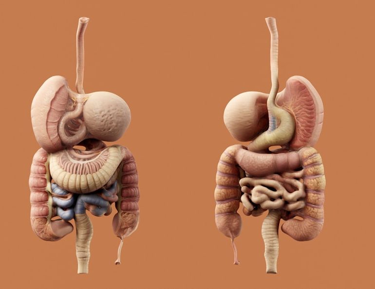
Before diving into diseases and treatments, let’s get acquainted with the organs that make up the urinary system and how they work together.
1.1 Kidneys – The Body’s Natural Filters
- Function: Remove waste products, excess salts, and water from the blood, producing urine.
- Key terms: Glomerulus, nephrons, renal pelvis.
- Health tip: Stay hydrated (≈2–3 L of water daily for most adults) to keep the filtration process efficient.
- Function: Two narrow muscular tubes that propel urine from each kidney to the bladder via peristaltic waves.
- Red flag: Sudden blockage (e.g., from a kidney stone) can cause severe flank pain—often called “renal colic.”
- Function: A muscular sac that expands to store urine until you’re ready to void.
- Important concept: Detrusor muscle contracts during urination; sphincter muscles relax to allow flow.
- Men: Approximately 20 cm long, passes through the prostate and penis.
- Women: About 4 cm long, ending just outside the vaginal opening.
- Why length matters: Women’s shorter urethra makes them more prone to UTIs.
- Prostate gland: Produces seminal fluid; can enlarge with age (benign prostatic hyperplasia, BPH).
- Testes, epididymis, vas deferens: Involved in sperm production and transport.
- What they are: Crystalline deposits (commonly calcium oxalate) that form in the kidneys and may travel down the ureters.
- Warning signs: Sudden, severe flank pain radiating to groin, hematuria (blood in urine), nausea, vomiting.
- Risk factors: Dehydration, high sodium diet, excessive animal protein, certain metabolic disorders (e.g., hyperparathyroidism).
- Diagnostic tools: Non‑contrast CT scan (gold standard), ultrasound, or plain X‑ray (KUB).
- Treatment options:
- Who gets it? Men over 50; prevalence rises to ~70% by age 80.
- Symptoms (often remembered by the acronym “LUTS”):
- Diagnostic work‑up: Digital rectal exam (DRE), PSA test (to rule out cancer), uroflowmetry, and possibly a bladder ultrasound.
- Medical management:
- Surgical options: Transurethral resection of the prostate (TURP) remains the gold standard; newer minimally invasive therapies include laser enucleation and prostatic urethral lift.
- Definition: Urgency with or without urge incontinence, often accompanied by frequency and nocturia.
- Causes: Detrusor muscle overactivity, neurologic disease, bladder outlet obstruction.
- First‑line therapy: Lifestyle modifications (fluid management, bladder training, pelvic floor exercises).
- Pharmacologic options: Antimuscarinics (oxybutynin, tolterodine) or β3‑agonists (mirabegron).
- Advanced treatments: Onabotulinum toxin A injections, sacral neuromodulation, peripheral tibial nerve stimulation.
- Prevalence: 2nd most common cancer in men worldwide.
- Screening: PSA test + digital rectal exam; MRI and biopsy for definitive diagnosis.
- Risk stratification: Gleason score, PSA level, tumor stage.
- Management pathways:
- Types: Stress UI (leakage with coughing/sneezing), urge UI (leakage after sudden urge), mixed UI, overflow UI.
- Evaluation: Physical exam, bladder stress test, urodynamic studies if needed.
- Conservative care: Pelvic floor muscle training (Kegel exercises), weight loss, smoking cessation.
- Medical options: Duloxetine (for stress UI), antimuscarinics (for urge UI).
- Surgical interventions: Mid‑urethral sling, artificial urinary sphincter, bulking agents.
- What it reveals: Infection, blood, protein, crystals, pH, specific gravity.
- When used: Initial assessment of UTIs, hematuria work‑up, monitoring after stone passage.
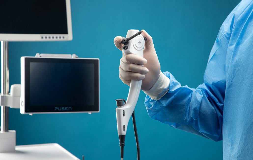
- Cystoscopy: Direct visualization of bladder and urethra; essential for tumor detection, stone retrieval, or evaluating strictures.
- Ureteroscopy: Miniaturized scope passed up the ureter; used for stone fragmentation and biopsies.
- Purpose: Assess bladder storage and emptying pressures; crucial for complex incontinence or neurogenic bladder.
- Components: Cystometry, pressure‑flow study, electromyography.
- Serum Creatinine & eGFR: Kidney function.
- PSA (Prostate‑Specific Antigen): Prostate cancer screening.
- Blood electrolytes: Evaluate metabolic causes of stone formation (e.g., hypercalcemia).
- Alpha‑blockers (tamsulosin) – Quick relief for BPH and aid stone passage.
- 5‑alpha‑reductase inhibitors (finasteride) – Long‑term prostate shrinkage.
- Mirabegron – β3‑agonist for OAB with fewer dry‑mouth side effects than antimuscarinics.
- SGLT2 inhibitors – Originally for diabetes, now shown to reduce risk of kidney stone formation by altering urinary calcium excretion.
- Focal Therapy for Prostate Cancer: High‑Intensity Focused Ultrasound (HIFU) and Cryotherapy target only cancerous tissue, preserving surrounding structures.
- Artificial Intelligence (AI) in Imaging: AI‑driven MRI interpretation improves detection of clinically significant prostate cancer, reducing unnecessary biopsies.
- Gene‑Based Treatments: PARP inhibitors (olaparib) for patients with BRCA mutations; immunotherapy (pembrolizumab) for MSI‑high tumors.
- Use a pill organizer (daily or weekly) to avoid missed doses.
- Set reminders on your phone for timing-sensitive drugs (e.g., tamsulosin taken 30 min after dinner).
- Track side effects in a journal; report persistent issues to your doctor promptly.
- Kidney health: Aim for 2–3 L of water daily; add a splash of lemon for citrate, which can inhibit stone formation.
- Prostate health: Incorporate foods rich in lycopene (tomatoes, watermelon) and omega‑3 fatty acids (salmon, flaxseed).
- Bladder health: Limit carbonated drinks, artificial sweeteners, and acidic foods if you have OAB.
- Kegel exercises: Contract pelvic floor muscles for 5 seconds, relax 5 seconds; repeat 10–15 times, three times a day.
- Aerobic activity: 150 minutes/week of moderate exercise improves circulation, reduces BPH symptoms, and supports overall kidney function.
- UTI prevention: Carry a portable urinalysis strip kit if you’re prone to infections; empty bladder regularly on long trips.
- Stone watch: When flying, stay hydrated and avoid excessive caffeine/alcohol; consider a low‑oxalate snack pack.
- Prostate cancer follow‑up: Keep a digital copy of pathology reports and imaging; schedule annual PSA tests as advised.
- Counseling: Cognitive‑behavioral therapy (CBT) helps manage anxiety related to incontinence or frequent bathroom trips.
- Support groups: Online forums (e.g., Kidney Stone Community, Prostate Cancer Support) provide peer encouragement and practical tips.
1.2 Ureters – The Transport Tubes
1.3 Bladder – The Storage Reservoir
1.4 Urethra – The Exit Pathway
1.5 Male Reproductive Organs (Often Included in Urology)
Takeaway: Knowing the basic anatomy helps you recognize symptoms early—whether it’s a burning sensation during urination (possible urethral irritation) or lower back pain (potential kidney involvement).
—
2. Common Urologic Conditions – Symptoms, Causes, and When to Seek Help
Urology covers a broad spectrum of disorders. Below are the most frequently encountered conditions, presented with actionable advice for each.
2.1 Urinary Tract Infections (UTIs)
| Aspect | Details |
|——–|———|
| Typical symptoms | Frequent urge to urinate, burning sensation, cloudy or foul‑smelling urine, pelvic discomfort. |
| Risk factors | Female anatomy, sexual activity, pregnancy, catheter use, diabetes, incomplete bladder emptying. |
| Diagnosis | Urine dipstick → culture if recurrent. |
| First‑line treatment | 3–7 days of antibiotics (e.g., nitrofurantoin, trimethoprim‑sulfamethoxazole). |
| Prevention tips | • Stay hydrated.
• Urinate before/after intercourse.
• Wipe front‑to‑back.
• Avoid prolonged bladder retention. |
When to call a urologist: Recurrent UTIs (≥3 in a year), kidney involvement (fever, flank pain), or persistent symptoms despite antibiotics.
2.2 Kidney Stones (Nephrolithiasis)
1. Conservative – Increased fluid intake, pain control (NSAIDs, opioids if needed).
2. Medical expulsive therapy – Alpha‑blockers (tamsulosin) to relax ureteral smooth muscle.
3. Procedural – Shock wave lithotripsy (SWL), ureteroscopy with laser lithotripsy, percutaneous nephrolithotomy (PCNL) for large stones.
Actionable tip: Aim for a urine output of >2 L/day; consider a diet lower in oxalate-rich foods (spinach, nuts) if you’re prone to stones.
2.3 Benign Prostatic Hyperplasia (BPH)
– Lower urinary tract symptoms: weak stream, hesitancy, dribbling.
– Urinary urgency and frequency, especially nocturia (nighttime trips).
– Alpha‑blockers (tamsulosin, alfuzosin) – relax prostate smooth muscle.
– 5‑alpha‑reductase inhibitors (finasteride, dutasteride) – shrink prostate size over months.
Practical advice: Limit caffeine and alcohol, practice timed voiding, and keep a bladder diary to track symptom patterns.
2.4 Overactive Bladder (OAB)
Quick tip: Keep a bladder diary for at least three days; it helps both you and your urologist pinpoint triggers.
2.5 Prostate Cancer
– Active surveillance for low‑risk disease.
– Radical prostatectomy, radiation therapy, brachytherapy, or hormone therapy for intermediate/high‑risk disease.
– Emerging options: Focal therapy (HIFU, cryotherapy), immunotherapy, PARP inhibitors for specific genetic mutations.
Action point: Discuss PSA testing with your primary care physician, especially if you have a family history or are over 50.
2.6 Urinary Incontinence (UI)
Takeaway: Early intervention often prevents progression; never accept “it’s just part of aging.”
—
3. Diagnostic Tools – How Urologists Find the Root Cause
Modern urology blends classic physical examinations with high‑tech imaging and laboratory studies. Understanding these tools can demystify the process and reduce anxiety.
3.1 Urinalysis & Urine Culture
3.2 Imaging Modalities
| Modality | Best For | Pros | Cons |
|———-|———-|——|——|
| Ultrasound | Kidney size, hydronephrosis, bladder volume | No radiation, bedside use | Operator dependent, limited stone detection |
| Non‑contrast CT | Kidney stones, complex anatomy | Highest sensitivity for stones | Radiation exposure |
| MRI (multiparametric) | Prostate cancer staging, soft‑tissue detail | No radiation, excellent soft‑tissue contrast | Cost, limited availability |
| X‑ray (KUB) | Radiopaque stones, foreign bodies | Quick, inexpensive | Misses radiolucent stones |
3.3 Endoscopic Procedures
3.4 Urodynamic Testing
3.5 Laboratory Markers
Bottom line: A combination of history, physical exam, and targeted diagnostics leads to precise treatment plans. Don’t hesitate to ask your urologist why a particular test is recommended.
—
4. Treatment Landscape – From Lifestyle Tweaks to Cutting‑Edge Technology
Urology is a field where lifestyle counseling sits side‑by‑side with robotic surgery. Below we outline the spectrum of interventions, emphasizing actionable steps you can start today.
4.1 Lifestyle & Preventive Strategies
| Issue | Lifestyle Change | Expected Impact |
|——-|——————|—————–|
| Kidney stones | Increase water intake, limit salt & animal protein, moderate oxalate foods | Reduces stone recurrence by up to 50% |
| BPH | Regular aerobic exercise, weight control, limit caffeine/alcohol | Improves urinary flow and nocturia |
| UTI | Probiotic use (Lactobacillus), cranberry extracts (mixed evidence) | May lower recurrence in women |
| OAB | Bladder training (timed voiding), limit bladder irritants (caffeine, spicy foods) | Decreases urgency episodes |
4.2 Pharmacologic Advances
4.3 Minimally Invasive Procedures
| Procedure | Indication | Key Benefits |
|———–|————|————–|
| Shock Wave Lithotripsy (SWL) | Small‑ to medium‑size kidney stones | Outpatient, no incision |
| Ureteroscopic laser lithotripsy | Mid‑ureter stones, larger stones | High stone‑free rates |
| Transurethral Resection of the Prostate (TURP) | Moderate BPH | Durable symptom relief |
| Laser Prostate Enucleation (HoLEP) | Large prostates | Less bleeding, shorter catheter time |
| Robotic‑assisted radical prostatectomy | Localized prostate cancer | Precise nerve‑sparing, quicker recovery |
4.4 Emerging Technologies
Action step: If you’re facing a major decision (e.g., surgery vs. active surveillance), ask your urologist about the latest minimally invasive options and whether you qualify for clinical trials.
—
5. Living with a Urologic Condition – Practical Tips for Daily Life
A diagnosis is just the beginning. Managing symptoms, medication schedules, and follow‑up appointments can feel overwhelming. Here are evidence‑based strategies to keep you in control.
5.1 Medication Management
5.2 Nutrition & Hydration
5.3 Exercise & Pelvic Floor Strengthening
5.4 Travel and Lifestyle Adjustments
5.5 Mental Health Considerations
Living with chronic urinary symptoms can affect confidence and social life.
Remember: You are not alone, and many resources exist to help you thrive despite a urologic diagnosis.
—
Conclusion – Key Takeaways for a Healthier Urinary System
1. Know your anatomy – Recognizing how kidneys, ureters, bladder, and urethra function empowers you to spot early warning signs.
2. Act on symptoms promptly – Burning urination, flank pain, or changes in urinary flow deserve timely evaluation to prevent complications.
3. Leverage diagnostics wisely – Urinalysis, imaging, and endoscopy are tools that guide precise treatment; never hesitate to ask why a test is recommended.
4. Combine lifestyle with medical care – Hydration, diet, pelvic floor exercises, and weight management complement medications and procedures for optimal outcomes.
5. Stay informed about modern treatments – From shock wave lithotripsy to robotic prostate surgery and AI‑assisted imaging, today’s urologic care offers less invasive, more effective options.
6. Prioritize follow‑up and mental well‑being – Regular appointments, medication adherence, and emotional support are essential components of long‑term urinary health.
By integrating these insights into your daily routine, you can maintain a robust urinary system, reduce the risk of common urologic conditions, and navigate any necessary treatments with confidence. Your kidneys, bladder, and prostate are vital partners in overall wellness—treat them with the respect and care they deserve.
—
If you found this guide helpful, share it with friends or family members who might benefit. For personalized advice, schedule a consultation with a board‑certified urologist who can tailor a plan to your unique health profile.

At King Edward Medical College, he was the recipient of the “Most Distinguished Student” award and graduated at the top of his class. Upon completing his medical education, Dr. Zafar went on to pursue his urology training at Royal Shrewsbury Hospital and at Ealing and Hammersmith Hospitals in England. In 1994, he completed a Fellowship in Endourology at Methodist Hospital in Indianapolis, Indiana. The focus of his fellowship was in urinary and bladder stones, male and female incontinence, and male impotence.
Dr. Zafar has been an active member of the American Urological Association, American Association of Clinical Urologists, and Society of Laparoscopic Surgeons.