

—
Introduction – Why This Topic Can’t Be Ignored
Imagine waking up in the middle of the night, rushing to the bathroom, only to discover you simply can’t pee. The panic, the discomfort, the lingering fear that something is seriously wrong – it’s a scenario many people would rather never experience. Yet, urinary retention—the inability to completely empty the bladder—is far more common than you might think.
According to recent health‑care data, up to 7 % of men over 50 and a smaller but still significant percentage of women will encounter acute or chronic urinary retention at some point in their lives. If left untreated, this condition can lead to infections, bladder damage, kidney problems, and a sharp decline in quality of life.
That’s why this guide dives deep into everything you need to know: what urinary retention really is, why it happens, how to spot it early, and—most importantly—what you can do about it. By the end, you’ll have a clear, actionable roadmap to protect your bladder health and seek the right care when needed.
—
1. What Is Urinary Retention? (And How Does It Differ From “Just Holding It In”?)
1.1 Defining the Condition
Urinary retention is the incomplete emptying of the bladder despite the desire to urinate. It can be categorized as:
| Type | Description | Typical Onset |
|——|————-|—————|
| Acute retention | Sudden, painful inability to urinate; often a medical emergency. | Minutes to hours |
| Chronic retention | Gradual, often painless reduction in urine flow; bladder may stretch over time. | Weeks to months |
| Partial vs. complete | Partial: some urine passes, but a significant amount remains. Complete: no urine passes at all. | — |
1.2 The Bladder’s Role in the Urinary System
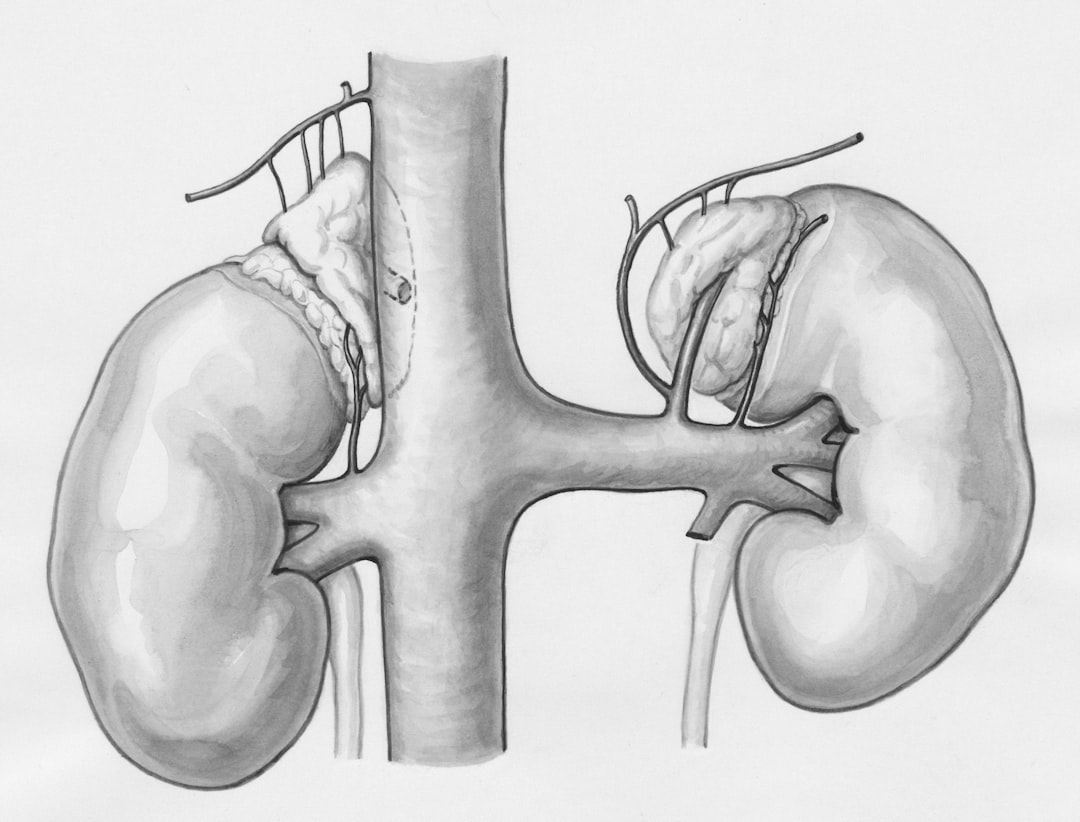
To appreciate why retention occurs, think of the bladder as a flexible reservoir that fills with urine from the kidneys via the ureters. Two muscle groups orchestrate emptying:
1. Detrusor muscle – contracts to push urine out.
2. Internal/external sphincters – relax to allow flow, then contract to stop it.
When either the muscle’s contractility or the sphincter’s coordination is disrupted, urine can get “stuck,” leading to retention.
1.3 Common Misconceptions
| Myth | Reality |
|——|———-|
| “It’s just me holding my pee.” | Holding urine occasionally is normal; retention is a physiological failure to empty, not a voluntary act. |
| “Only older men get it.” | While men over 50 are at higher risk (often due to prostate issues), women, children, and younger adults can develop retention for many reasons. |
| “If I can’t pee, I need a catheter right away.” | Acute retention often requires immediate catheterization, but chronic cases may be managed with medication, lifestyle changes, or intermittent self‑catheterization. |
—
2. The Root Causes – Why Does Urinary Retention Happen?
Understanding the underlying trigger is essential for effective treatment. Below are the most frequent culprits, grouped by anatomical, neurological, pharmacological, and functional categories.
2.1 Anatomical Blockages
| Cause | How It Leads to Retention | Typical Population |
|——-|—————————|——————–|
| Benign Prostatic Hyperplasia (BPH) | Enlarged prostate compresses the urethra, impeding flow. | Men >50 |
| Urethral stricture | Scar tissue narrows the urethra. | Men with prior catheter use, infections, or trauma |
| Pelvic organ prolapse | In women, uterus or bladder descent can kink the urethra. | Post‑menopausal women |
| Bladder stones / tumors | Physical obstruction inside the bladder. | All ages (stones more common in men) |
2.2 Neurological Disorders
The bladder is a neuro‑muscular organ; any disruption in nerve signals can cause retention.
| Condition | Mechanism | Symptoms Beyond Retention |
|———–|———–|—————————|
| Multiple sclerosis (MS) | Demyelination interferes with detrusor contraction. | Numbness, vision problems |
| Spinal cord injury | Interrupts reflex arcs between brain and bladder. | Paralysis, loss of sensation |
| Parkinson’s disease | Impaired dopamine pathways affect sphincter coordination. | Tremor, rigidity |
| Diabetic neuropathy | Chronic high glucose damages autonomic nerves. | Tingling, foot ulcers |
2.3 Medications & Substances
Certain drugs relax the bladder’s detrusor muscle or tighten the sphincter.
| Drug Class | Examples | Why It Causes Retention |
|————|———-|————————|
| Anticholinergics | Oxybutynin, Tolterodine | Block acetylcholine → weak detrusor |
| Opioids | Morphine, Oxycodone | Central nervous system depressant → reduced urge |
| Alpha‑blockers (paradoxical effect) | Prazosin (high dose) | Over‑relax sphincter → urinary stasis |
| Antidepressants (TCAs) | Amitriptyline | Anticholinergic side‑effects |
| Antihistamines | Diphenhydramine | Anticholinergic properties |
2.4 Functional & Lifestyle Triggers
Even without a structural or neurological problem, habits can tip the balance.
- Chronic constipation – Pressure on the bladder and urethra.
- Heavy lifting or straining – Increases intra‑abdominal pressure, temporarily compressing the urethra.
- Dehydration – Concentrated urine irritates the bladder, leading to spasms and incomplete emptying.
- Post‑surgical swelling – Particularly after pelvic or prostate surgery.
- Sudden onset after trauma or surgery
- Accompanied by fever, chills, or flank pain (possible infection)
- Persistent inability to urinate for > 6 hours (risk of bladder rupture)
- Nighttime waking to urinate (nocturia) – may indicate chronic retention.
- Dribbling after finishing – sign of sphincter dysfunction.
- Lower back or flank pain – urine backing up toward kidneys.
- Unexplained swelling of the lower abdomen – a full bladder can mimic a mass.
- What: Patient inserts a sterile catheter several times a day to fully empty the bladder.
- When to use: Chronic retention with high residual volumes but intact bladder function.
- Advantages: Low infection risk when done correctly, preserves bladder tone, no permanent hardware.
- Key tip: Use a single‑use catheter or a clean technique with a reusable catheter; rotate insertion sites to avoid urethral trauma.
- Goal: Improve coordination between detrusor and sphincter.
- Methods: Timed voiding, urge suppression techniques, biofeedback, and Kegel exercises (especially in women).
- Evidence: Studies show up to 30 % reduction in residual volumes after 12 weeks of structured training.
2.5 Red Flags: When Retention Signals Something Serious
If any of these appear, seek emergency medical care immediately.
—
3. Spotting the Signs – Symptoms & When to Get Tested
Early detection can prevent complications such as urinary tract infections (UTIs), bladder stones, or kidney damage. Below is a checklist of the most common (and some subtle) symptoms.
3.1 Classic Symptoms
| Symptom | What It Means |
|———|—————-|
| Weak or intermittent stream | Detrusor not contracting strongly |
| Feeling of incomplete emptying | Residual urine remains after voiding |
| Frequent urge to urinate but little output | Overactive bladder trying to compensate |
| Abdominal or suprapubic pain | Bladder stretching beyond capacity |
| No urine output despite full bladder | Acute retention – medical emergency |
3.2 Subtle Clues
3.3 Diagnostic Tools
| Test | What It Shows | Typical Use |
|——|—————|————-|
| Post‑void residual (PVR) ultrasound | Volume of urine left after voiding | First‑line for chronic retention |
| Uroflowmetry | Flow rate and pattern | Detects obstruction vs. weak detrusor |
| Cystoscopy | Direct visual of bladder & urethra | Identifies strictures, stones, tumors |
| Urodynamic studies | Comprehensive pressure‑flow analysis | Complex neurogenic cases |
| Blood work (creatinine, BUN) | Kidney function | Rule out renal impairment from back‑pressure |
| CT or MRI | Structural abnormalities in pelvis/spine | Post‑trauma, suspected tumors |
A post‑void residual volume > 100 mL generally signals clinically significant retention. Values above 300 mL often require intervention.
—
4. Treatment Options – From Lifestyle Tweaks to Surgical Solutions
The best approach depends on type (acute vs. chronic), cause, severity, and patient preferences. Below we break down the spectrum of interventions, highlighting the pros, cons, and typical success rates.
4.1 Immediate Management of Acute Retention
| Step | Action | Reason |
|——|——–|——–|
| Catheterization | Insert a straight (in‑and‑out) catheter or indwelling Foley if prolonged drainage needed. | Relieves bladder pressure, prevents damage. |
| Pain control | NSAIDs or mild opioids (short‑term) | Reduces discomfort and spasm. |
| Identify trigger | Review meds, recent surgery, trauma. | Prevent recurrence. |
| Follow‑up | Usually within 24–48 h for urology review. | Determine long‑term plan. |
4.2 Pharmacologic Therapies for Chronic Retention
| Medication | Mechanism | Typical Candidates |
|————|———–|——————–|
| Alpha‑blockers (tamsulosin, alfuzosin) | Relax smooth muscle in prostate & bladder neck → lower outlet resistance. | Men with BPH‑related retention. |
| 5‑alpha‑reductase inhibitors (finasteride, dutasteride) | Shrink prostate over months. | Moderate‑to‑severe BPH, combined with alpha‑blocker. |
| Cholinergic agents (bethanechol) | Stimulate detrusor contraction. | Neurogenic retention, post‑operative cases. |
| Botulinum toxin A (Botox) injections | Reduces overactive sphincter tone. | Detrusor‑sphincter dyssynergia (often in spinal cord injury). |
| Antibiotics | Treat concurrent UTI. | When infection present; not a primary retention cure. |
Success tip: If you’re on an anticholinergic for overactive bladder, discuss alternatives with your doctor—these meds can paradoxically worsen retention.
4.3 Non‑Surgical Interventions
#### 4.3.1 Intermittent Self‑Catheterization (ISC)
#### 4.3.2 Bladder Training & Pelvic Floor Therapy
#### 4.3.3 Lifestyle Modifications
| Change | Why It Helps |
|——–|————–|
| Increase fluid intake (1.5–2 L/day) | Prevents concentrated urine, reduces irritation. |
| Limit caffeine & alcohol | Both act as diuretics and bladder irritants. |
| High‑fiber diet | Reduces constipation‑related pressure on bladder. |
| Weight management | Excess abdominal fat can compress the pelvis. |
| Timed voiding (every 3–4 h) | Prevents over‑distension of bladder. |
4.4 Surgical Options – When the Blockade Is Too Strong
| Procedure | Indication | How It Works | Recovery |
|———–|————|————–|———-|
| Transurethral Resection of the Prostate (TURP) | BPH causing severe obstruction | Removes prostate tissue via a resectoscope. | Hospital stay 1–2 days; catheter 1 week. |
| Urethral dilation or internal urethrotomy | Short urethral stricture | Mechanical widening of the urethra. | Outpatient; may need repeat. |
| Suprapubic catheter placement | Long‑term drainage when urethral catheter not tolerated. | Catheter placed through lower abdomen into bladder. | Requires surgical insertion; care similar to Foley. |
| Artificial urinary sphincter (AUS) or male sling | Post‑prostatectomy incontinence with retention | Mechanical device that compresses urethra when needed. | 1–2 weeks hospital stay; device lifespan 5–10 years. |
| Bladder augmentation (enterocystoplasty) | Small, poorly compliant bladder (rare) | Uses intestinal segment to enlarge bladder capacity. | Major abdominal surgery; 2–3 weeks hospitalization. |
Choosing surgery should involve a thorough discussion of **benefits, risks, and

At King Edward Medical College, he was the recipient of the “Most Distinguished Student” award and graduated at the top of his class. Upon completing his medical education, Dr. Zafar went on to pursue his urology training at Royal Shrewsbury Hospital and at Ealing and Hammersmith Hospitals in England. In 1994, he completed a Fellowship in Endourology at Methodist Hospital in Indianapolis, Indiana. The focus of his fellowship was in urinary and bladder stones, male and female incontinence, and male impotence.
Dr. Zafar has been an active member of the American Urological Association, American Association of Clinical Urologists, and Society of Laparoscopic Surgeons.